. 2013 Oct 24;8(10):e77660.
doi: 10.1371/journal.pone.0077660. eCollection 2013.

Identification of a core bacterial community within the large intestine of the horse

Affiliations

Identification of a core bacterial community within the large intestine of the horse

Kirsty Dougal et al. PLoS One. .

Abstract

The horse has a rich and complex microbial community within its gastrointestinal tract that plays a central role in both health and disease. The horse receives much of its dietary energy through microbial hydrolysis and fermentation of fiber predominantly in the large intestine/hindgut. The presence of a possible core bacterial community in the equine large intestine was investigated in this study. Samples were taken from the terminal ileum and 7 regions of the large intestine from ten animals, DNA extracted and the V1-V2 regions of 16SrDNA 454-pyrosequenced. A specific group of OTUs clustered in all ileal samples and a distinct and different signature existed for the proximal regions of the large intestine and the distal regions. A core group of bacterial families were identified in all gut regions with clear differences shown between the ileum and the various large intestine regions. The core in the ileum accounted for 32% of all sequences and comprised of only seven OTUs of varying abundance; the core in the large intestine was much smaller (5-15% of all sequences) with a much larger number of OTUs present but in low abundance. The most abundant member of the core community in the ileum was Lactobacillaceae, in the proximal large intestine the Lachnospiraceae and in the distal large intestine the Prevotellaceae. In conclusion, the presence of a core bacterial community in the large intestine of the horse that is made up of many low abundance OTUs may explain in part the susceptibility of horses to digestive upset.


Conflict of interest statement

Competing Interests: Patricia A. Harris is employed by one of the funders of this research (WALTHAM Centre for Pet Nutrition, Melton Mowbray, Leicestershire. LE14 4RT). The authors can confirm that they have adhered to all the PLOS ONE policies on sharing data and materials. The authors also confirm that none of the authors record a Conflict of Interest.

Figures

Figure 1
Figure 1. Phyla identified and relative proportion of each in different compartments of the horse’s large intestine.
Data shown from the Ileum, caecum, right ventral colon (RVC), left ventral colon (LVC), left dorsal colon (LDC), right dorsal colon (RDC), small colon and faeces. The category others includes; TM7, Tenericutes, synergistetes, Cyanobacteria/Chloroplast, SR1, Elusimicrobia, Fusobacteria, Chloroflexi.
Figure 2
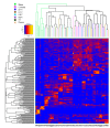
Figure 2. Heatmap showing OTUs in different compartments of the horse’s large intestine.
Data shown from the ileum (A) caecum (B), right ventral colon (C), left ventral colon (D), left dorsal colon (E), right dorsal colon (F), small colon (G) and faeces (H) found at 0.1% or greater abundance as a percentage of the total number of sequences (accounts for 49.28% of the total sequences). The cladogram on the vertical axis shows the phylogenetic relationship between the individual OTUs included in this analysis. The cladogram on the horizontal axis shows the phylogentic relationship between all the OTUs identified in each sample.
Figure 3
Figure 3. The core bacterial community in different compartments of the horse’s large intestine.
The core community is defined as those OTUs (clustered at 97% similarity) present in all ten animals and which abundances are 0.1% (or greater) of the total number of sequences in that gut region. The lower pale blue section of the bar indicates the proportion that is not part of the core. The remaining individual coloured sections represent each OTU of which the core is comprised; Ileum (7), caecum (31), right ventral colon (RVC) (33), left ventral colon (LVC) (28), left dorsal colon (LDC) (12), right dorsal colon (RDC) (18), small colon (16) and faeces (25). For further details see table S3.
Figure 4
Figure 4. Relative abundance of the most prevalent bacterial families identified as the core bacterial community.
Regions of the large intestine included are the caecum, right ventral colon (RVC), left ventral colon (LVC), left dorsal colon (LDC), right dorsal colon (RDC), small colon and faeces. Values presented do not account all for members of stated families found in each gut region only those identified as being part of the core; error bars represent the standard deviation. A-Lactobacillaceae B-Clostridiales_Insertae Sedis XIII C- Coriobacteriaceae D-Acidaminococcacaea E-Porphyromondaceae F-Spirochaetaceae G-Clostridiaceae 1 H-Erysipelotrichacea I-Fibrobacteraceae J-Ruminococcaceae K-Lachnospiraceae L-Prevotellaceae.

References

    1. Turnbaugh PJ, Gordon JI (2009) The core gut microbiome, energy balance and obesity. J Physiol 587: 4153-4158. doi:10.1113/jphysiol.2009.174136. PubMed: 19491241. - DOI - PMC - PubMed
    1. Turnbaugh PJ, Quince C, Faith JJ, McHardy AC, Yatsunenko T et al. (2010) Organismal, genetic, and transcriptional variation in the deeply sequenced gut microbiomes of identical twins. Proc Natl Acad Sci USA 107(16): 7503-7508. doi:10.1073/pnas.1002355107. PubMed: 20363958. - DOI - PMC - PubMed
    1. Sekelja M, Berget I, Næs T, Rudi K (2011) Unveiling an abundant core microbiota in the human adult colon by a phylogroup-independent searching approach. ISME J 5: 519-531. doi:10.1038/ismej.2010.129. PubMed: 20740026. - DOI - PMC - PubMed
    1. Jalanka-Tuovinen J, Salonen A, Nikkilä J, Immonen O, Kekkonen R et al. (2011) Intestinal microbiota in healthy adults: Temporal analysis reveals individual and common core and relation to intestinal symptoms. PLOS ONE 6(7): e23035. doi:10.1371/journal.pone.0023035. PubMed: 21829582. - DOI - PMC - PubMed
    1. Shade A, Handelsman J (2012) Beyond the venn diagram: the hunt for a core microbiome. Environ Microbiol 14(1): 4-12. doi:10.1111/j.1462-2920.2011.02585.x. PubMed: 22004523. - DOI - PubMed

Publication types

LinkOut - more resources