. 2013 Oct 30;8(10):e77749.
doi: 10.1371/journal.pone.0077749. eCollection 2013.

Calcium/calcineurin synergizes with prostratin to promote NF-κB dependent activation of latent HIV

Affiliations

Calcium/calcineurin synergizes with prostratin to promote NF-κB dependent activation of latent HIV

Jonathan K Chan et al. PLoS One. .

Abstract

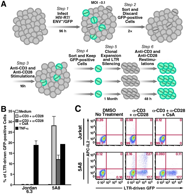
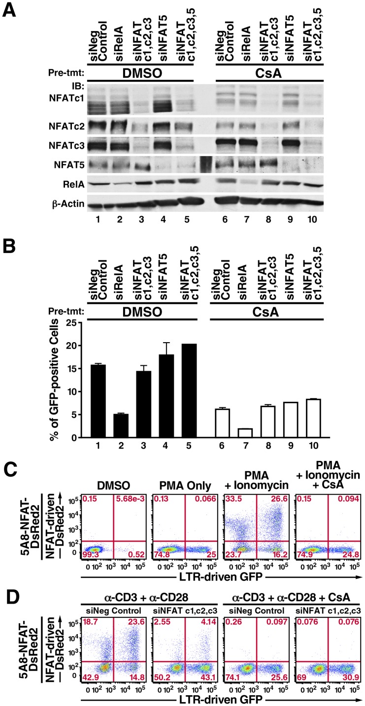
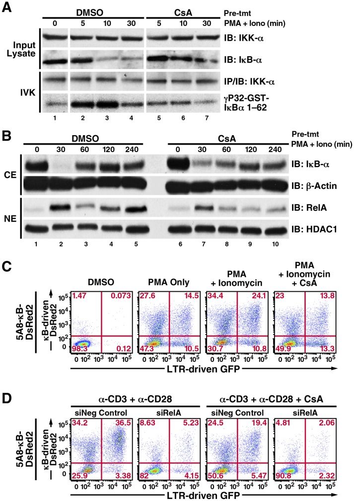
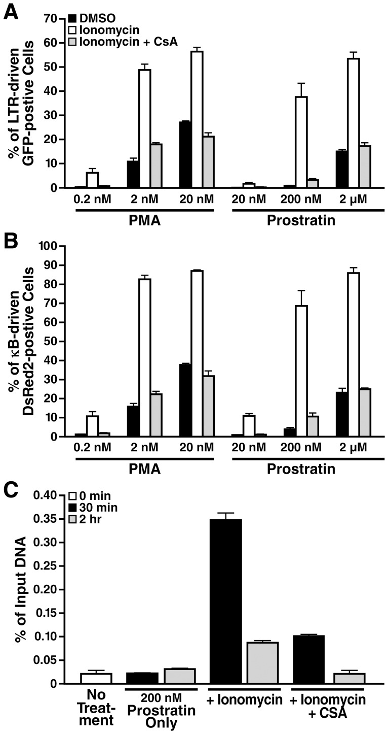
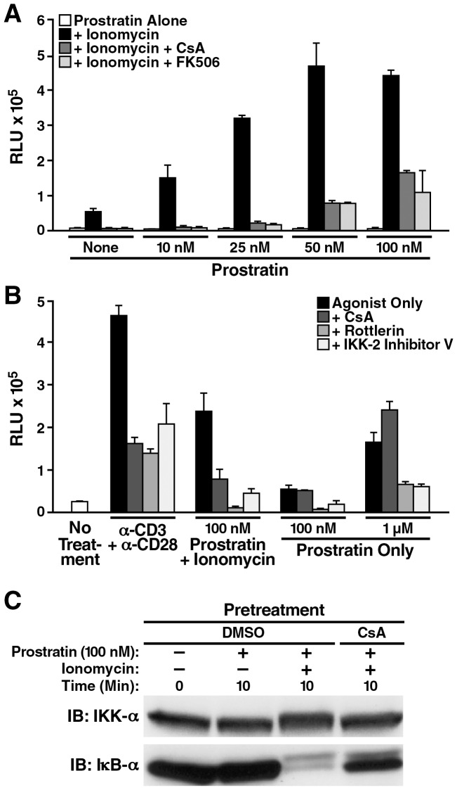
Attempts to eradicate HIV have been thwarted by the persistence of a small pool of quiescent memory CD4 T cells that harbor a transcriptionally silent, integrated form of the virus that can produce infectious virions following an anamnestic immune response. Transcription factors downstream of T-cell receptor activation, such as NF-κB/Rel and nuclear factor of activated T cells (NFAT) transcription members, are considered important regulators of HIV transcription during acute HIV infection. We now report studies exploring their precise role as antagonists of HIV latency using cell and primary CD4 T cell models of HIV-1 latency. Surprisingly, RNA interference studies performed in J-Lat CD4 T cells suggested that none of the NFATs, including NFATc1, NFATc2, NFATc3, and NFAT5, played a key role in the reactivation of latent HIV. However, cyclosporin A markedly inhibited the reactivation response. These results were reconciled when calcium signaling through calcineurin was shown to potentiate prostratin induced activation of NF-κB that in turn stimulated the latent HIV long terminal repeat (LTR). Similar effects of calcineurin were confirmed in a primary CD4 T cell model of HIV latency. These findings highlight an important role for calcineurin in NF-κB-dependent induction of latent HIV transcription. Innovative approaches exploiting the synergistic actions of calcineurin and prostratin in the absence of generalized T-cell activation merit exploration as a means to attack the latent viral reservoir.


Conflict of interest statement

Competing Interests: The authors have declared that no competing interests exist.

Figures

Figure 1
Figure 1. 5A8 cells, a J-Lat cell line responsive to TCR stimulation and CsA inhibition.
A, generation of 5A8 cells. B, LTR-GFP reporter activity of Jordan J-Lat 6.3 and 5A8 cells in response to different stimuli in the presence or absence of CsA. Cells were treated with DMSO (white, light gray, black) or 500 nM CsA (dark gray) and then left untreated (white) or stimulated with platebound anti-CD3 (10 µg/ml) and soluble anti-CD28 antibodies (2 µg/ml) (light and dark gray) or 10 ng/ml TNF-α (black) for 24 h, followed by FACS analysis to determine the percentage of GFP-positive cells (y-axis). Values are mean ± SEM from three experiments. Crosslinking of TCR induced latent HIV reporter activity in 5A8 cells that was partially inhibited by CsA. These responses were not observed in Jordan J-Lat 6.3 cells. C, TCR-crosslinking-dependent induction of IL-2 in Jurkat and 5A8 cells. Cells were treated with DMSO (left and middle columns) or 500 nM CsA (right column) and incubated with Brefeldin A alone or together with anti-CD3 and anti-CD28 antibodies prepared as above for 16 h. Cells were collected, stained with anti-human IL-2 antibody conjugated to allophycocyanin (APC), and analyzed by FACS. 5A8 cells produced more IL-2 than their parental Jurkat cells, probably because they were selected by their enriched expression of TCR complexes. CsA abolished IL-2 production but only partially inhibited LTR-driven GFP reporter activity.
Figure 2
Figure 2. NFAT does not appear to be involved in latent HIV transcription in 5A8 cells.
A, immunoblotting analysis of RelA and NFAT knockdowns in 5A8 cells. Negative control siRNA (lanes 1 and 6), siRNAs against RelA (lanes 2 and 7), NFATc1, NFATc2, and NFATc3 (lanes 3 and 8), NFAT5 alone (lanes 4 and 9), and all four NFATs (lanes 5 and 10) were introduced into 5A8 cells by Amaxa nucleofection twice within 48 h. Cells were then treated with DMSO (lanes 1 to 5) or 500 nM CsA (lanes 6 to 10) for 2 h, stimulated with anti-CD3 and anti-CD28 antibodies for 24 h, and collected for immunoblotting or FACS analyses. CsA reduced the levels of NFATc1 and NFATc2, which autoamplified their expressions after TCR crosslinking. B, FACS analysis of LTR-driven GFP reporter activity (y-axis). Values are mean ± SEM from four experiments. Reporter activity remained partially sensitive to CsA inhibition (lanes 6 to 10) but was insensitive to NFAT knockdown (lanes 3 to 5). C, characterization of 5A8-NFAT-DsRed2 cells. Cells were pretreated with DMSO (panels 1–3) or 500 nM CsA for 2 h (panel 4) and incubated with 20 nM PMA (panel 2), or 20 nM PMA and 2 µM ionomycin (panels 3 and 4) for 24 h, followed by FACS analysis. PMA alone induced activity of the GFP reporter but not the DsRed2 reporter (panel 2). Ionomycin and PMA induced both reporters (panel 3). CsA fully inhibited combined PMA/ionomycin induction of DsRed2 reporter but only partially suppressed GFP reporter activity to levels similar to those after stimulation with PMA only (panel 4). D, effects of NFATc1, NFATc2 and NFATc3 knockdown on expression of NFAT-dependent DsRed2 and LTR-driven GFP. 5A8-NFAT-DsRed2 cells were nucleofected with negative control siRNA (panels 1 and 3) or siRNA against NFATc1, NFATc2, and NFATc3 (panels 2 and 4) twice within 48 h, treated with DMSO (panels 1 and 2) or 500 nM CsA (panels 3 and 4) for 2 h, and stimulated with anti-CD3 and anti-CD28 antibodies as in Figure 1. NFATc1, NFATc2 and NFATc3 knockdown decreased expression of the DsRed2 reporter but did not inhibit GFP reporter activity (panels 1 and 2). CsA partially reduced GFP reporter expression even in the absence of NFAT (panels 2 and 4).
Figure 3
Figure 3. CsA reduces PKC-induced NF-κB/RelA activation and LTR-transcription in 5A8 cells.
A, in vitro kinase assay of IκBα phosphorylation by IKK. 5A8 cells were pretreated with DMSO or 500 nM CsA for 2 h, stimulated with 20 nM PMA and 2 µM ionomycin for the indicated times, lysed, and in vitro kinase assays was performed using glutathione-S-transferase IκBα (1–62) as the substrate as described . CsA delayed the degradation of endogenous IκBα induced by PMA/ionomycin, which correlated with reduced phosphorylation of GST-IκBα by immunoprecipitated IKK-α. B, analysis of PMA/ionomycin-induced IκBα degradation and RelA nuclear translocation. 5A8 cells were treated with DMSO or 500 nM CsA for 2 h, stimulated with 20 nM PMA and 2 µM ionomycin, and fractionated into nuclear and cytoplasmic extracts. Immunoblotting analyses revealed that CsA interfered with complete degradation of cytoplasmic IκBα at 30 min and reduced its reappearance at 60 min. CsA also reduced the first and second rounds of nuclear NF-κB/RelA expression at 30 min and 120 min. C, characterization of 5A8-κB-DsRed2 cells. Cells were treated as in Figure 2C. Unlike 5A8-NFAT-DsRed2 cells, PMA alone induced both κB-dependent DsRed2 and LTR-driven GFP reporter activities (panel 2). Combined PMA/ionomycin stimulation further enhanced the activities of both reporters (panel 3), which were partially suppressed by CsA to levels similar to those after stimulation with PMA only (panel 4). D, effects of RelA knockdown on expression of κB-dependent DsRed2 and LTR-driven GFP. 5A8-κB-DsRed2 cells were nucleofected with negative control siRNA (panels 1 and 3) or siRNA against RelA (panels 2 and 4) twice within 48 h, followed by drug and antibody treatments as in Figure 2d. RelA knockdown suppressed both κB-DsRed2 and LTR-driven GFP reporter activities (panels 2 and 4).
Figure 4
Figure 4. Calcium/calcineurin signaling synergizes with prostratin to antagonize latent HIV-1 by targeting RelA to LTR.
A and B, dose-response analyses of LTR-driven GFP and κB-driven DsRed2 reporter activities. 5A8 cells were pretreated with DMSO (black and white bars) or 500 nM CsA (gray bars) for 2 h, followed by stimulation with PMA or prostratin only (black bars), or together with 2 µM ionomycin (white and gray bars) for 24 h at the concentration of the phorbol ester as indicated before FACS analysis. Values are mean ± standard deviation from one representative experiment. CsA potently suppressed the synergistic effect of ionomycin with PMA or prostratin and reverted back the activities of DsRed2 and GFP reporters to PMA- or prostratin-only levels. C, ChIP analysis of RelA recruitment to HIV-1 LTR. 5A8 cells were pretreated with DMSO or 500 nM CsA, followed by stimulation with 200 nM prostratin alone or together with 2 µM ionomycin for times indicated. Cells were fixed and subjected to chromatin immunoprecipitation with anti-RelA antibodies. Enrichment of LTR-chromatin in anti-RelA immunoprecipitates was expressed as a percentage of input chromatin. Ionomycin synergized with suboptimal dosage of prostratin to promote robust to substantial RelA occupancy at HIV-1 LTR at 30 min and 2 h, respectively. CsA effectively diminished RelA recruitment.
Figure 5
Figure 5. PKC/IKK inhibition strongly reduces prostratin/ionomycin-induced latent HIV transcription in primary CD4+ T cells.
A and B, evaluating inhibitors of relevant pathway in suppressing NL4-3 luciferase reporter activities in latently infected primary CD4+ T cells. A, latently infected cells pretreated with DMSO (white and black bars), calcineurin inhibitors CsA (dark gray bar) or FK-506 (light gray bar) were incubated in medium that contained no or suboptimal dosages of prostratin (0 to 100 nM, white bars), or contained both prostratin and 1.5 µM ionomycin (black, dark and light gray bars) for 48 h. Either calcineurin inhibitors inhibited the dose-dependent synergistic effect between suboptimal dosages of prostratin and ionomycin on luciferase reporter activities. B, latently infected cells pretreated with DMSO (white and black bars) or various kinase/phosphatase inhibitors (dark, medium and light gray bars) were incubated in media alone (white bar), or media containing anti-CD3/anti-CD28 Dynabeads (1∶1 ratio), or prostratin, in the presence or absence of 1.5 µM ionomycin as indicated for 30 h. All inhibitors suppressed reporter activities induced by TCR crosslinking or prostratin/ionomycin, which involved induction of calcium/calcineurin signaling, but CsA was ineffective against reporter activities induced by prostratin alone. In both A and B, the RLU were normalized based on total protein present in the various cell lysates. All stimulations were performed in triplicate with error bars representing ± standard deviation. Results are representative of experiments performed with cells from three independent donors. C, analysis of prostratin/ionomycin-induced IκBα degradation in primary CD4 cells. PBMC-purified CD4 cells were treated with DMSO or 500 nM CsA for 2 h, stimulated with a suboptimal dose of prostratin at 100 nM in the presence or absence of 1.5 µM ionomycin for 10 min and whole-cell lysate was prepared. Immunoblotting analyses revealed that CsA effectively reduced the effect of stimulus-coupled degradation of cytoplasmic IκBα.

References

    1. Prins JM, Jurriaans S, van Praag RM, Blaak H, van Rij R, et al. (1999) Immuno-activation with anti-CD3 and recombinant human IL-2 in HIV-1-infected patients on potent antiretroviral therapy. AIDS 13: 2405–2410. - PubMed
    1. Stellbrink HJ, van Lunzen J, Westby M, O’Sullivan E, Schneider C, et al. (2002) Effects of interleukin-2 plus highly active antiretroviral therapy on HIV-1 replication and proviral DNA (COSMIC trial). AIDS 16: 1479–1487. - PubMed
    1. Reuse S, Calao M, Kabeya K, Guiguen A, Gatot JS, et al. (2009) Synergistic activation of HIV-1 expression by deacetylase inhibitors and prostratin: implications for treatment of latent infection. PLoS One 4: e6093. - PMC - PubMed
    1. Yang HC, Shen L, Siliciano RF, Pomerantz JL (2009) Isolation of a cellular factor that can reactivate latent HIV-1 without T cell activation. Proc Natl Acad Sci U S A 106: 6321–6326. - PMC - PubMed
    1. Yang HC, Xing S, Shan L, O'Connell K, Dinoso J, et al. (2009) Small-molecule screening using a human primary cell model of HIV latency identifies compounds that reverse latency without cellular activation. J Clin Invest 119: 3473–3486. - PMC - PubMed

Publication types

MeSH terms