. 2013 Oct 28;8(10):e78061.
doi: 10.1371/journal.pone.0078061. eCollection 2013.

Aqueous extract of Bambusae Caulis in Taeniam inhibits PMA-induced tumor cell invasion and pulmonary metastasis: suppression of NF-κB activation through ROS signaling

Affiliations

Aqueous extract of Bambusae Caulis in Taeniam inhibits PMA-induced tumor cell invasion and pulmonary metastasis: suppression of NF-κB activation through ROS signaling

Aeyung Kim et al. PLoS One. .

Abstract

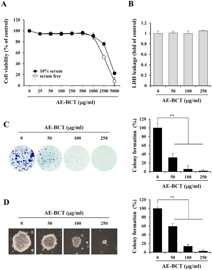
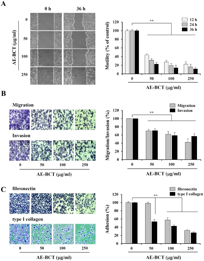
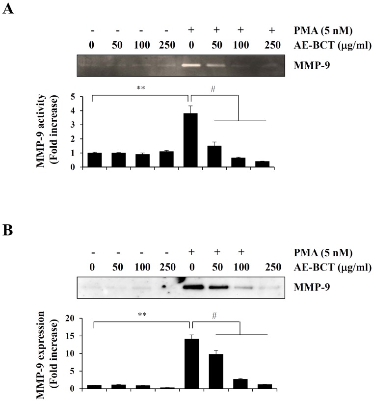
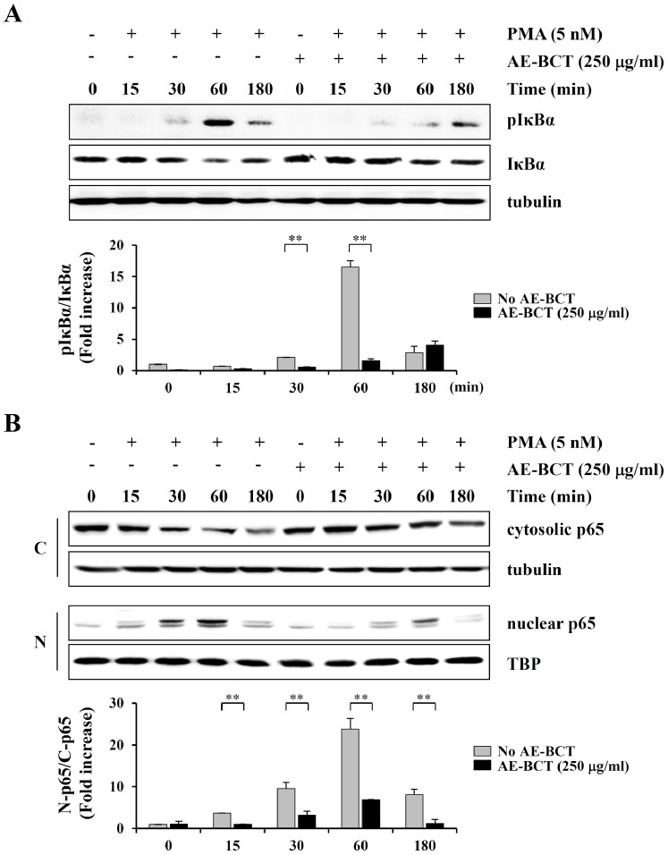
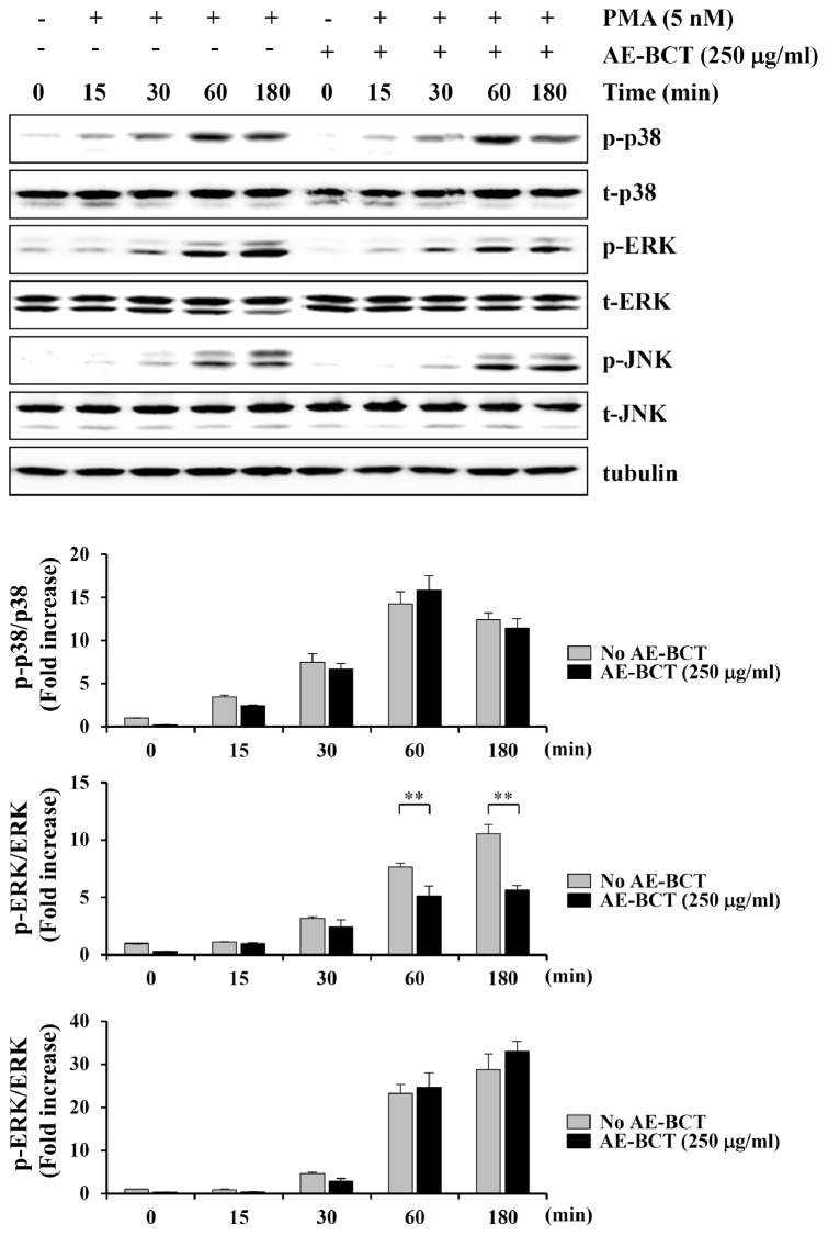
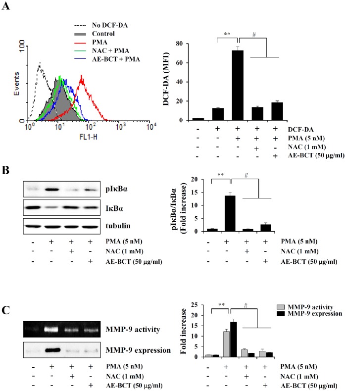
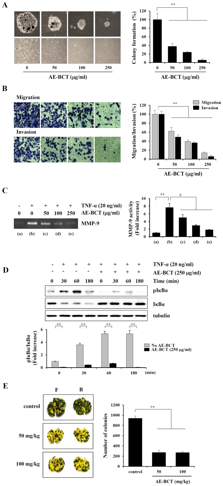
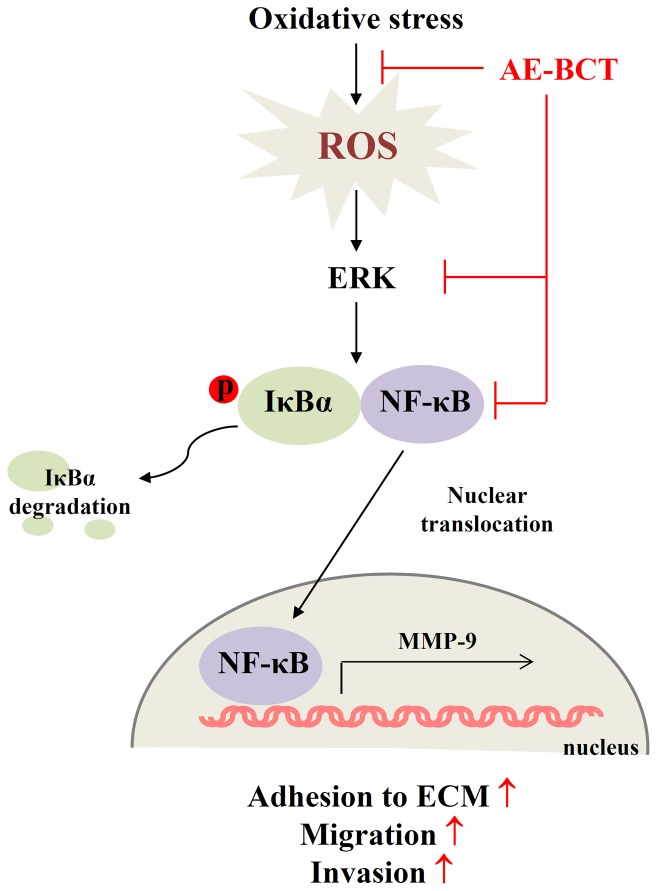
Bamboo shavings (Bambusae Caulis in Taeniam, BCT) are widely used as a traditional Chinese medicine to control hypertension and cardiovascular disease, and to alleviate fever, vomiting, and diarrhea. It has been demonstrated that BCT reduces ovalbumin-induced airway inflammation by regulating pro-inflammatory cytokines, and decreases tumor growth in tumor-bearing mice. However, the effects of BCT on the metastatic potential of malignant cancer cells and the detailed mechanism of its anti-metastatic activity have not been examined previously. In this study, we investigated whether an aqueous extract of BCT (AE-BCT) reduces the metastatic potential of HT1080 cells, and elucidated the underlying anti-metastatic mechanism. In addition, we examined whether AE-BCT administration inhibits pulmonary metastasis of intravenously injected B16F10 cells in C57BL/6J mice. AE-BCT (50-250 µg/ml) dose-dependently suppressed colony-forming activity under anchorage-dependent and -independent growth conditions. Pretreatment with AE-BCT efficiently inhibited cell migration, invasion, and adhesion. AE-BCT also dramatically suppressed PMA-induced MMP-9 activity and expression by blocking NF-κB activation and ERK phosphorylation. Production of intracellular ROS, a key regulator of NF-κB-induced MMP-9 activity, was almost completely blocked by pretreatment with AE-BCT. Furthermore, daily oral administration of AE-BCT at doses of 50 and 100 mg/kg efficiently inhibited lung metastasis of B16F10 cells injected into the tail veins of C57BL/6J mice with no systemic toxicity. These results demonstrate that AE-BCT significantly reduced the metastatic activity of highly malignant cancer cells by suppressing MMP-9 activity via inhibition of ROS-mediated NF-κB activation. These results indicate that AE-BCT may be a safe natural product for treatment of metastatic cancer.


Conflict of interest statement

Competing Interests: The authors have declared that no competing interests exist.

Figures

Figure 1
Figure 1. Effect of AE-BCT on the viability and colony-forming activity of HT1080 cells.
Cells seeded onto a 96-well culture plate were treated with 25 to 5000 µg/mL AE-BCT with or without serum for 48 h. Cell cytotoxicity was estimated by MTT (A) and LDH release (B) assays. (C) Representative photographs of anchorage-dependent colony formation in the presence or absence of AE-BCT. At the end of incubation, cells were stained with crystal violet. The areas of 20 representative colonies were measured, and relative colony formation was quantitated using ImageJ. (D) Effect of AE-BCT on anchorage-independent colony formation. After 2 weeks of incubation, colonies on soft agar were observed and the diameters of 20 representative colonies were measured. Data are expressed as means ± SD. **p<0.01 vs untreated control.
Figure 2
Figure 2. Effect of AE-BCT on the in vitro migration, invasion, and adhesion to ECM of HT1080 cells.
(A) Confluent cell monolayers were treated with mitomycin C (25 µg/mL) for 1 h and an injury line was drawn. Cell debris was removed by washing and the cells were incubated in medium containing 10% FBS with or without 50, 100, or 250 µg/mL AE-BCT. Wound migration was monitored using a phase-contrast microscope (×40) at the indicated time points and the relative width of the gap region was quantified by measuring four selected fields. (B) Cells pre-incubated for 12 h with or without AE-BCT were harvested and suspended in serum-free DMEM at a density of 1×10⁶ cells/mL. After filling the upper Transwell chamber with cells suspended in serum-free DMEM (1×10⁵ cells/100 µL) and the lower chamber with 10% FBS/DMEM (650 µL), the cells were incubated. Cells that migrated and invaded to the lower surface of the membrane were stained and observed using a phase-contrast microscope. (C) Cells pre-incubated for 12 h with or without AE-BCT were harvested and suspended in serum-free DMEM at a density of 5×10⁵ cells/mL. After seeding on FN- or collagen-coated wells (1×10⁵ cells/well/200 µL), cells were incubated for 1 h and then washed to remove unattached cells. Attached cells were quantified by crystal violet staining. The relative degrees of migration, invasion, and adhesion to ECM were quantified using ImageJ. Data are expressed as means ± SD of three independent experiments. **p<0.01 vs untreated control.
Figure 3
Figure 3. Effects of AE-BCT on MMP-9 activity and expression in HT1080 cells.
Cells pre-treated with AE-BCT for 12 h were incubated in serum-free medium with or without 5 nM PMA for a further 24 h. Conditioned media were collected and analyzed for the activity and expression of MMP-9 by gelatin zymography (A) and Western blotting (B), respectively. Bar graphs shows fold increases in band intensity compared with untreated control cells. Data are expressed as means ± SD of three independent experiments. **p<0.01 vs untreated control, # p<0.01 vs PMA stimulation.
Figure 4
Figure 4. Effect of AE-BCT on NF-κB activation in HT1080 cells.
(A) Control and AE-BCT-pre-treated HT1080 cells were stimulated with 5 nM PMA for the indicated periods of time and cell lysates were subjected to Western blotting of the phosphorylation and degradation of IκBα. (B) To examine the nuclear translocation of the NF-κB p65 subunit in response to PMA stimulation, cell lysates were fractionated into cytosolic and nuclear compartments, and subjected to Western blotting. After normalization to α-tubulin or TBP expression, relative ratios of pIκBα/IκBα and nuclear p65/cytosolic p65 were determined. Data are expressed as means ± SD of two independent experiments. **p<0.01 vs no AE-BCT.
Figure 5
Figure 5. Effect of AE-BCT on MAPK activation in HT1080 cells.
Control and AE-BCT-pre-treated HT1080 cells were stimulated with 5 nM PMA for the indicated period of time and cell lysates were subjected to Western blotting of the phosphorylation of p38, ERK, and JNK. Relative ratios of phosphorylated forms to total levels were determined after normalization to α-tubulin expression. Data are expressed as means ± SD of two independent experiments. **p<0.01 vs no AE-BCT.
Figure 6
Figure 6. Effect of AE-BCT on intracellular ROS generation, and involvement of ROS in NF-κB activation and MMP-9 activity.
(A) Cells pre-treated with NAC (1 mM) for 1 h or AE-BCT (50 µg/ml) for 12 h were stimulated with PMA (5 nM) for 3 h, incubated with DCF-DA (5 µM) for 30 min at 37°C, and then subjected to flow cytometry assay of ROS levels. Data are expressed as means ± SD of three independent experiments. **p<0.01 vs untreated control, # p<0.01 vs PMA stimulation (B) Cells pre-treated with NAC or AE-BCT were stimulated with PMA (5 nM) for 60 min, and pIκBα and IκBα protein levels were determined by Western blotting. (C) Conditioned media collected after 24 h of PMA stimulation were analyzed for MMP-9 activity and expression. Values are expressed as fold increases in band intensity compared with untreated control cells. Data are expressed as means ± SD of two independent experiments. **p<0.01 vs untreated control, # p<0.01 vs PMA stimulation.
Figure 7
Figure 7. Effect of AE-BCT on the in vitro metasatic potential and in vivo pulmonary metastasis of B16F10 cells.
(A) To examine anchorage-independent cell growth, a soft agar colony formation assay was performed. After 10 days of incubation with or without AE-BCT, colonies were observed (upper ×200, lower ×40) and the diameters of 20 representative colonies were measured. (B) B16F10 cells pre-treated with or without AE-BCT for 12 h were examined for Transwell migration and invasion capabilities. Data are expressed as means ± SD of two independent experiments. **p<0.01 vs untreated control (C) Cells pre-treated with AE-BCT for 12 h were incubated in serum-free medium with 20 ng/mL TNF-α for a further 24 h. Conditioned media were collected and analyzed for the activity of MMP-9 by gelatin zymography. **p<0.01 vs untreated control, # p<0.01 vs TNF-α stimulation (D) Control and AE-BCT-pre-treated B16F10 cells were stimulated with 20 ng/ml TNF-α for the 30, 60, and 180 min and cell lysates were examined for the phosphorylation and degradation of IκBα. After normalization to α-tubulin expression, relative ratios of pIκBα/IκBα were determined. Data are expressed as means ± SD of two independent experiments. **p<0.01 vs no AE-BCT (E) Cells (3×10⁵/200 µL PBS) were injected into the tail veins of 6-week-old female C57BL/6J mice, to which AE-BCT (50 or 100 mg/kg) was administered daily. At day 17, mice were killed and the black colonies on the lung surface were enumerated macroscopically. Images of metastatic lung nodules at the front (F) and back (B) are shown. Colonies were enumerated and relative inhibition compared with the control group was calculated. Data are representative of two independent experiments (n = 5 per group). **p<0.01 vs saline control.
Figure 8
Figure 8. Scheme of the anti-metastatic mechanism of AE-BCT.
AE-BCT significantly reduces the metastatic potential of malignant cancer cells by suppression of MMP-9 activity via inhibition of ROS-mediated NF-κB activation.

References

    1. Liotta LA, Steeg PS, Stetler-Stevenson WG (1991) Cancer metastasis and angiogenesis: an imbalance of positive and negative regulation. Cell 64: 327–336. - PubMed
    1. Chambers AF, Groom AC, MacDonald IC (2002) Dissemination and growth of cancer cells in metastatic sites. Nat Rev Cancer 2: 563–572. - PubMed
    1. Patel LR, Camacho DF, Shiozawa Y, Pienta KJ, Taichman RS (2011) Mechanisms of cancer cell metastasis to the bone: a multistep process. Future Oncol 7: 1285–1297. - PMC - PubMed
    1. Jinga DC, Blidaru A, Condrea I, Ardeleanu C, Dragomir C, et al. (2006) MMP-9 and MMP-2 gelatinases and TIMP-1 and TIMP-2 inhibitors in breast cancer: correlations with prognostic factors. J Cell Mol Med 10: 499–510. - PMC - PubMed
    1. Kim TS, Kim YB (1999) Correlation between expression of matrix metalloproteinase-2 (MMP-2), and matrix metalloproteinase-9 (MMP-9) and angiogenesis in colorectal adenocarcinoma. J Korean Med Sci 14: 263–270. - PMC - PubMed

Publication types

MeSH terms