. 2013 Oct 21;8(10):e78076.
doi: 10.1371/journal.pone.0078076. eCollection 2013.

Adult-specific systemic over-expression reveals novel in vivo effects of the soluble forms of ActRIIA, ActRIIB and BMPRII

Affiliations

Adult-specific systemic over-expression reveals novel in vivo effects of the soluble forms of ActRIIA, ActRIIB and BMPRII

Kengo Yamawaki et al. PLoS One. .

Abstract

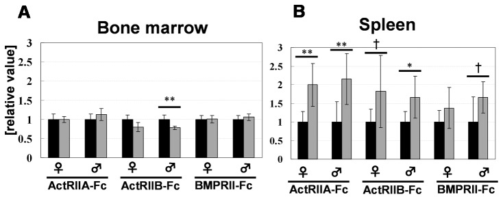
Bone morphogenetic proteins (BMPs)/growth differentiation factors (GDFs), which belong to the TGF-beta superfamily, are pleiotropic factors that play a role in regulating the embryonic development and postnatal homeostasis of various organs and tissues by controlling cellular differentiation, proliferation and apoptosis. Conventional transgenic and knockout (KO) mouse approaches have provided only limited information regarding the in vivo functions of BMP signaling in adult animals due to the effects on prenatal development and the difficulty in manipulating multiligand signals simultaneously. We recently produced transgenic chimeric mice(Tg chimeras) in which the soluble IgG1-Fc fusion protein of three BMP type II receptors (ActRIIA, ActRIIB, BMPRII) was highly circulated (281-709 μg/ml), specifically in adult mouse blood. Since each BMP receptor can bind to multiple BMP ligands, these Tg chimeras should be useful to investigate the effects of trapping multiple BMP ligands. Remarkably, some phenotypes were unexpected based on previous studies, such as KO mouse analyses, presumably representing the effects of the multiple ligand trapping. These phenotypes included increased red blood cells (RBCs) and decreased viability in adults. In a further study, we focused on the phenotype of increased RBCs and found that extramedullary hematopoiesis in the spleen, not in the bone marrow, was increased using histological and flow cytometric analyses. Although it remains to be elucidated whether the transgene products affect the tissues directly or indirectly, our data provide novel and important insight into the biological functions of the soluble IgG1-Fc fusion protein of three BMP type II receptors in adults, and our approach should have broad applications to research on other ligand receptor families and studies involving mouse models.


Conflict of interest statement

Competing Interests: The authors have the following interests. Financial support for this study was provided by Kyowa-Hakko Kirin Co. Ltd., the employer of Kengo Yamawaki, Tsutomu Okada, Takeshi Oshima, Makoto Kakitani and Kazuma Tomizuka. There are no patents, products in development or marketed products to declare. This does not alter the authors' adherence to all the PLOS ONE policies on sharing data and materials, as detailed online in the guide for authors.

Figures

Figure 1
Figure 1. Serum protein levels produced from the transgenes in each Tg chimeras.
A. Serum hEPO concentrations of the four types of eight-week-old Tg chimeras identified using a combination of two different promoters (A: used in our previous report, B: newly developed for this study) and the existence of puro and neo selection markers. ***, P<0.001 (Student’s t-test). Significant differences were observed in both females and males. B. The serum concentration of each soluble protein (ActRIIA-Fc, ActRIIB-Fc and BMPRII-Fc) in 11- to 16-week-old Tg chimeras was measured using ELISA (see the Materials and Methods). In the control chimeras, the Fc protein was not detectable. As described in Materials and Methods, the “control” chimeras are produced using ES cells in which the expression unit is not introduced.
Figure 2
Figure 2. Survival rate of each Tg chimeras.
The term before 10 weeks was abbreviated because all chimeric mice were still alive.
Figure 3
Figure 3. Summary and grouping of the phenotypes observed in each Tg chimeras (11- to 16 weeks old).
A. Venn diagram representing the common phenotypes of the three Tg chimeras. The ellipses represent the phenotypes obtained from each Tg chimeras. The common phenotypes are shown in the overlapping sections. B. Summary of the phenotypes. The notes correspond to the data shown in Figure 3A. For example, VII means that the features were common to all three (ActRIIA-Fc, ActRIIB-Fc and BMPRII-Fc) Tg chimeras.
Figure 4
Figure 4. Histological images of the spleen in each group of Tg chimeras (8 weeks old).
A. Control, B. ActRIIA-Fc, C. ActRIIB-Fc and D. BMPRII-Fc. As shown in the magnified images, increased extramedullary erythrocytic hematopoiesis was observed. The arrowheads represent foci of erythroblastic islets. Such foci were observed in all three Tg chimeras at a very high frequency (shown as percentages in the figure) but not in the control spleens (only one of the 10 mice exhibited a less severe phenotype). Each scale bar indicates 200 μm.
Figure 5
Figure 5. Increased extramedullary hematopoiesis was observed in the chimeric mouse spleens.
Results of the flow cytometric analysis of the bone marrow (A) and spleen (B). The black bar indicates the control chimeras and the gray bar indicates the Tg chimeras. Samples were obtained from 8-week-old Tg chimeras (N=6). Each experiment was performed independently, and a control was prepared for each procedure. The ratio of [% of gated cells] in each Tg chimera to that observed in the control chimeras was described as [relative value]. *, P<0.05 ; **, P<0.01; †, tendency (0.05t-test).

References

    1. Feng XH, Derynck R (2005) Specificity and versatility in tgf-beta signaling through Smads. Annu Rev Cell Dev Biol 21: 659-693. doi:10.1146/annurev.cellbio.21.022404.142018. PubMed: 16212511. - DOI - PubMed
    1. Massagué J (1998) TGF-beta signal transduction. Annu Rev Biochem 67: 753-791. doi:10.1146/annurev.biochem.67.1.753. PubMed: 9759503. - DOI - PubMed
    1. Chang H, Brown CW, Matzuk MM (2002) Genetic analysis of the mammalian transforming growth factor-beta superfamily. Endocr Rev 23: 787-823. doi:10.1210/er.2002-0003. PubMed: 12466190. - DOI - PubMed
    1. Zhao GQ (2003) Consequences of knocking out BMP signaling in the mouse. Genesis 35: 43-56. doi:10.1002/gene.10167. PubMed: 12481298. - DOI - PubMed
    1. Kitisin K, Saha T, Blake T, Golestaneh N, Deng M et al. (2007) Tgf-beta signaling in development. Sci STKE 2007: cm1 PubMed: 17699101 - PubMed

Publication types

MeSH terms

Substances