. 2013 Oct 25;8(10):e78309.
doi: 10.1371/journal.pone.0078309. eCollection 2013.

A quantitative study of inhibitory interneurons in laminae I-III of the mouse spinal dorsal horn

Affiliations

A quantitative study of inhibitory interneurons in laminae I-III of the mouse spinal dorsal horn

Erika Polgár et al. PLoS One. .

Abstract

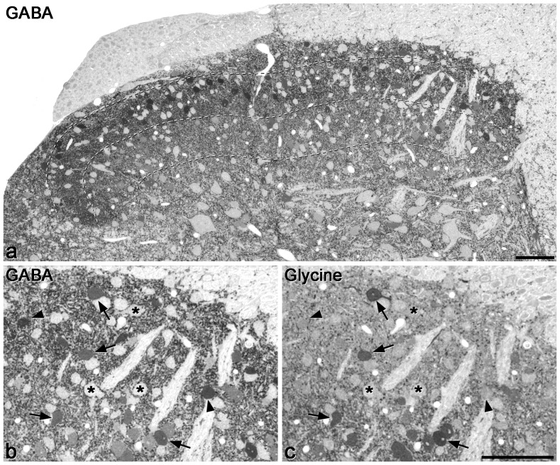
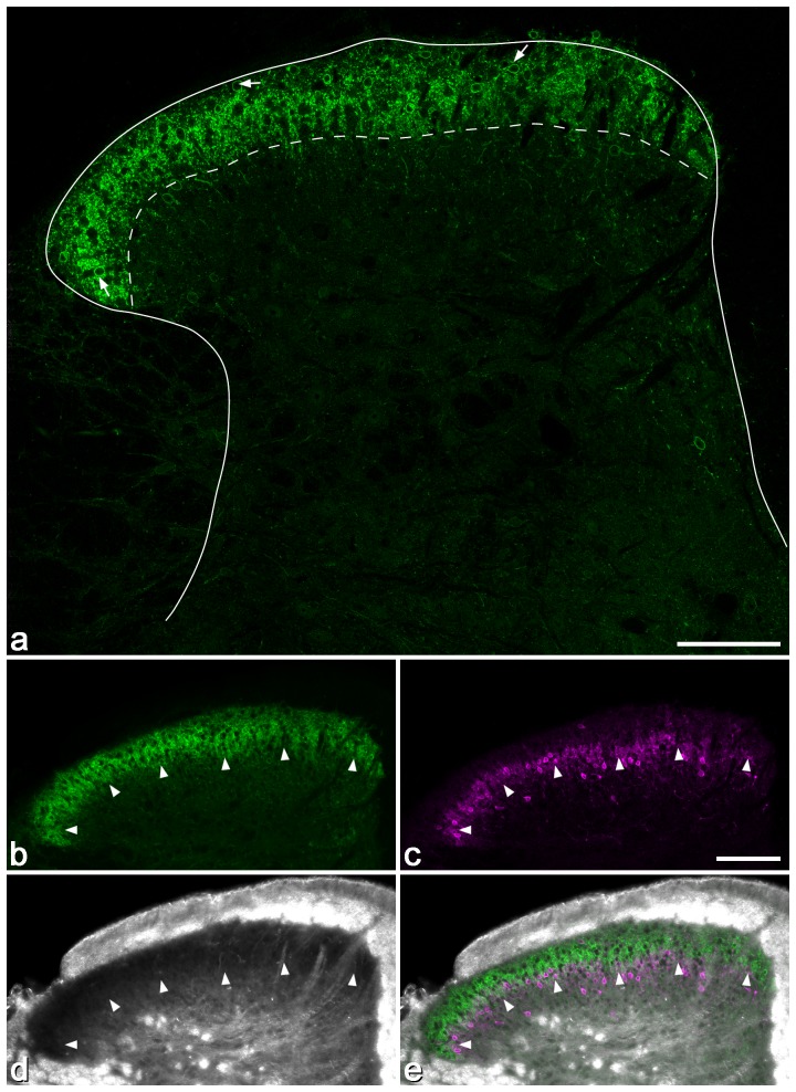
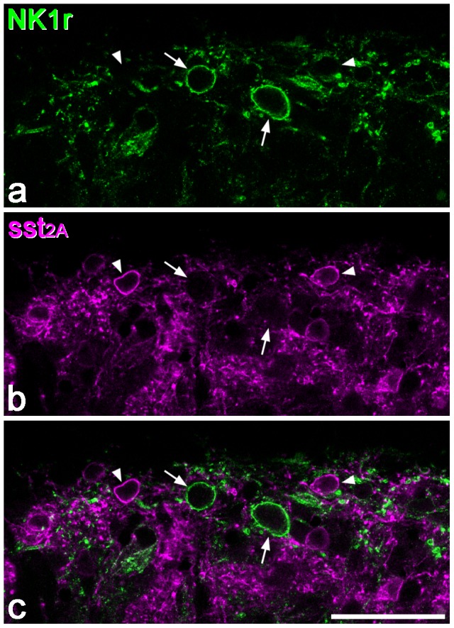
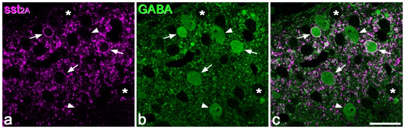
Laminae I-III of the spinal dorsal horn contain many inhibitory interneurons that use GABA and/or glycine as a neurotransmitter. Distinct neurochemical populations can be recognised among these cells, and these populations are likely to have differing roles in inhibiting pain or itch. Quantitative studies in rat have shown that inhibitory interneurons account for 25-40% of all neurons in this region. The sst2A receptor is expressed by around half the inhibitory interneurons in laminae I-II, and is associated with particular neurochemically-defined populations. Although much of the work on spinal pain mechanisms has been performed on rat, the mouse is now increasingly used as a model, due to the availability of genetically altered lines. However, quantitative information on the arrangement of interneurons is lacking in the mouse, and it is possible that there are significant species differences in neuronal organisation. In this study, we show that as in the rat, nearly all neurons in laminae I-III that are enriched with glycine also contain GABA, which suggests that GABA-immunoreactivity can be used to identify inhibitory interneurons in this region. These cells account for 26% of the neurons in laminae I-II and 38% of those in lamina III. As in the rat, the sst2A receptor is only expressed by inhibitory interneurons in laminae I-II, and is present on just over half (54%) of these cells. Antibody against the neurokinin 1 receptor was used to define lamina I, and we found that although the receptor was concentrated in this lamina, it was expressed by many fewer cells than in the rat. By estimating the total numbers of neurons in each of these laminae in the L4 segment of the mouse, we show that there are around half as many neurons in each lamina as are present in the corresponding segment of the rat.


Conflict of interest statement

Competing Interests: The authors have declared that no competing interests exist.

Figures

Figure 1
Figure 1. GABA and glycine immunoreactivity in a 0.5 µm thick transverse section of the mouse dorsal horn.
a shows the whole mediolateral extent of the dorsal horn stained for GABA, while b and c show the medial part of the dorsal horn at higher magnification, in the same section and in a section reacted for glycine, respectively. Many GABA-immunoreactive cell bodies are scattered throughout the dorsal horn, and although these vary in intensity, they are clearly darker than the immunonegative cells, some of which are marked with asterisks in b. The dark staining in the neuropil represents GABAergic axons and dendrites. Dashed lines show the ventral borders of laminae I, II and III. b and c are serial semithin sections, so the same cells are visible. A few cells with relatively strong glycine immunoreactivity are visible (some marked with arrows) and these are also GABA-immunoreactive. In addition, there are cells that are GABA- but not glycine-immunoreactive (two marked with arrowheads). Scale bars  = 100 µm.
Figure 2
Figure 2. Distribution of sst2A immunoreactivity in the mouse dorsal horn.
a: There is a dense band of immunostaining in laminae I and II, with much lower levels elsewhere in the grey matter. Immunoreactive cells (some marked with arrows) are scattered throughout laminae I and II, and present at much lower density in deeper laminae. The solid line shows the grey-white matter boundary and the dashed line the border between laminae II and III. b–f: The relationship between sst2A (green), PKCγ (magenta) and the appearance of the dorsal horn with dark-field illumination (DF). Note that although scattered PKCγ cells are present in lamina III, the ventral border of the PKCγ plexus (arrowheads) is at the same level as the ventral edge of sst2A staining, and that this corresponds to the lamina II/III border as seen with dark-field microscopy. All confocal images are from single optical sections. Scale bar  = 100 µm.
Figure 3
Figure 3. Confocal images showing sst2A-immunoreactive neurons in laminae I-II of the mouse dorsal horn in a transverse section used for quantitative analysis.
a: Part of the section scanned to reveal sst2A (green). Several immunoreactive cells are visible, and three of these are marked with arrows. b: The same field scanned to reveal NeuN (blue) and the nuclear stain DAPI (red). c: In the merged image, the dorsal border of the dorsal horn is shown with a solid line and the boundary between laminae II and III with a dashed line. In this image, neuronal nuclei appear magenta, while the nuclei of non-neuronal cells are red. Note that most neurons in laminae I-II are not sst2A-immunoreactive. The images were obtained from a single optical section. Scale bar  = 50 µm.
Figure 4
Figure 4. NK1r and sst2A in superficial dorsal horn.
Confocal image showing a single optical section through part of laminae I and IIo, scanned to reveal NK1r (green) and sst2A (magenta). a: Two NK1r-immunoreactive cell bodies (arrows) are visible in lamina I, surrounded by a plexus of labelled dendrites. b: Several sst2A-immunoreactive cells are visible in the same field (two shown with arrowheads). c: The merged image shows that none of these cells is labelled with both antibodies. Scale bar  = 50 µm.
Figure 5
Figure 5. Restriction of sst2A-immunoreactivity to GABAergic neurons in the superficial dorsal horn.
a: A confocal scan (single optical section) from the top surface of a Vibratome section that had been reacted to reveal sst2A (magenta) and GABA (green). Three sst2A-immunoreactive cell bodies are marked with arrows, and there are many other labelled profiles, most of which are dendrites cut in cross section. b: The same field scanned to reveal GABA. Several GABA-immunoreactive cell bodies can be seen and some of these are indicated with arrows or arrowheads. Many cell bodies that are not GABA-immunoreactive are also present, and two are marked with asterisks. c: The merged image reveals that the sst2A-immunoreactive cells are all immunostained for GABA, but that several GABA+ cells do not possess sst2A (two are indicated with arrowheads). Scale bar  = 20 µm.

References

    1. Polgár E, Hughes DI, Riddell JS, Maxwell DJ, Puskár Z, et al. (2003) Selective loss of spinal GABAergic or glycinergic neurons is not necessary for development of thermal hyperalgesia in the chronic constriction injury model of neuropathic pain. Pain 104: 229–239. - PubMed
    1. Todd AJ (2010) Neuronal circuitry for pain processing in the dorsal horn. Nat Rev Neurosci 11: 823–836. - PMC - PubMed
    1. Todd AJ, Sullivan AC (1990) Light microscope study of the coexistence of GABA-like and glycine-like immunoreactivities in the spinal cord of the rat. J Comp Neurol 296: 496–505. - PubMed
    1. Maxwell DJ, Rethelyi M (1987) Ultrastructure and synaptic connections of cutaneous afferent fibres in the spinal cord. Trends Neurosci 10: 117–123.
    1. Ribeiro-da-Silva A, Coimbra A (1982) Two types of synaptic glomeruli and their distribution in laminae I-III of the rat spinal cord. J Comp Neurol 209: 176–186. - PubMed

Publication types