. 2013 Oct 18;8(10):e78434.
doi: 10.1371/journal.pone.0078434. eCollection 2013.

Histamine from brain resident MAST cells promotes wakefulness and modulates behavioral states

Affiliations

Histamine from brain resident MAST cells promotes wakefulness and modulates behavioral states

Sachiko Chikahisa et al. PLoS One. .

Abstract

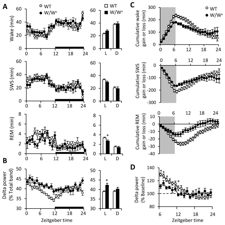
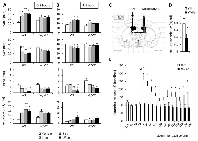
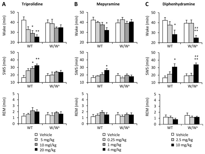
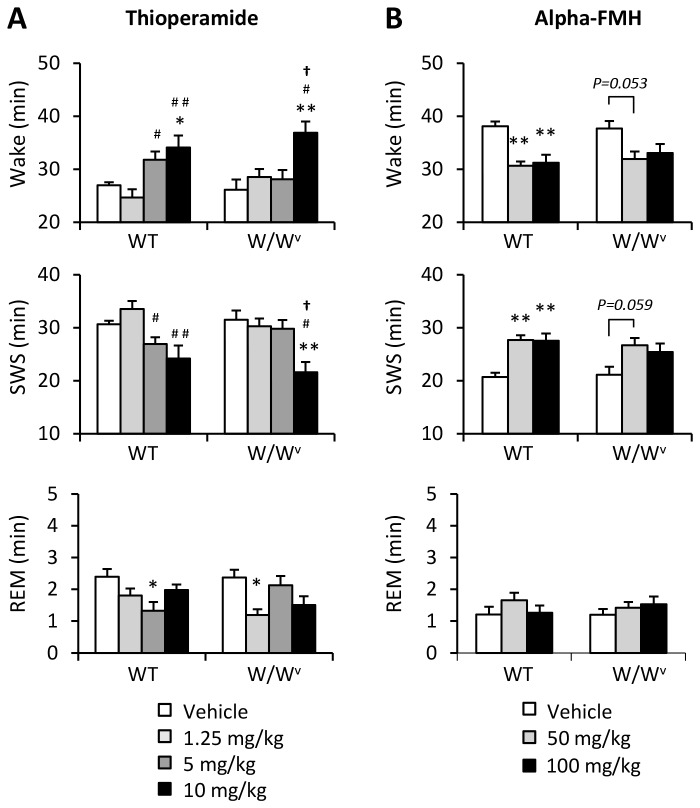
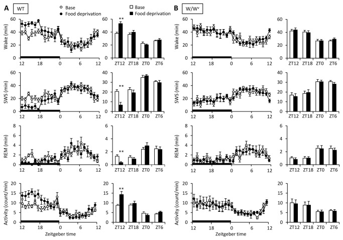
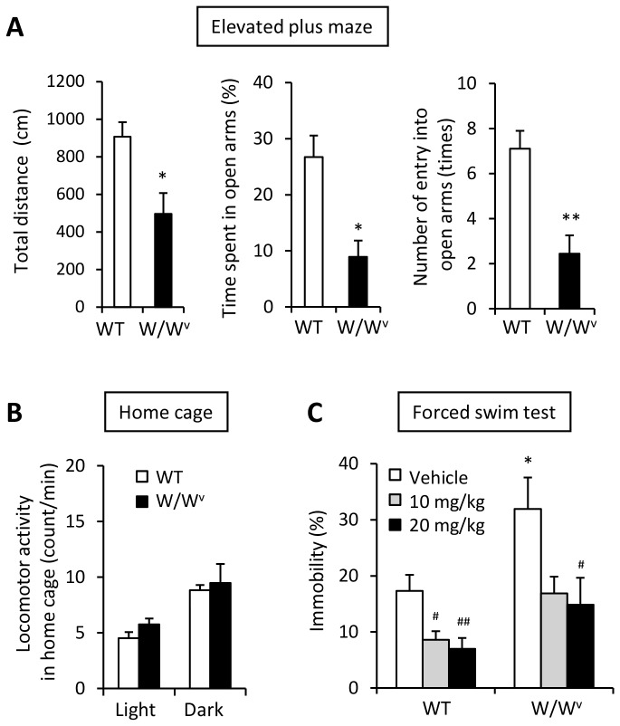
Mast cell activation and degranulation can result in the release of various chemical mediators, such as histamine and cytokines, which significantly affect sleep. Mast cells also exist in the central nervous system (CNS). Since up to 50% of histamine contents in the brain are from brain mast cells, mediators from brain mast cells may significantly influence sleep and other behaviors. In this study, we examined potential involvement of brain mast cells in sleep/wake regulations, focusing especially on the histaminergic system, using mast cell deficient (W/W(v)) mice. No significant difference was found in the basal amount of sleep/wake between W/W(v) mice and their wild-type littermates (WT), although W/W(v) mice showed increased EEG delta power and attenuated rebound response after sleep deprivation. Intracerebroventricular injection of compound 48/80, a histamine releaser from mast cells, significantly increased histamine levels in the ventricular region and enhanced wakefulness in WT mice, while it had no effect in W/W(v) mice. Injection of H1 antagonists (triprolidine and mepyramine) significantly increased the amounts of slow-wave sleep in WT mice, but not in W/W(v) mice. Most strikingly, the food-seeking behavior observed in WT mice during food deprivation was completely abolished in W/W(v) mice. W/W(v) mice also exhibited higher anxiety and depression levels compared to WT mice. Our findings suggest that histamine released from brain mast cells is wake-promoting, and emphasizes the physiological and pharmacological importance of brain mast cells in the regulation of sleep and fundamental neurobehavior.


Conflict of interest statement

Competing Interests: The authors have declared that no competing interests exist.

Figures

Figure 1
Figure 1. Baseline sleep/wake characterizations of WT and W/Wv mice.
(A) Time course for amount of wake (top) and SWS (middle) was not different between the two genotypes. Amount of REM (bottom) was slightly decreased in W/Wv mice during the light phase (L; ZT0-12) but was not during the dark phase (D; ZT12-24). (B) EEG delta power in SWS was significantly higher in W/Wv mice during the light phase (L; ZT0-12). The bar graphs indicate the averaged data for each 12-hour period across ZT0-12 (L) and ZT12-24 (D). (C) Cumulative sleep/wake loss and gain compared with baseline conditions for the sleep deprivation experiment. Sleep deprivation began at ZT0 and ended at ZT6 (the shadow areas). (D) The rebound response of EEG delta power in SWS after sleep deprivation was attenuated in W/Wv mice. Data is expressed as the percent change from the baseline value (dotted line) at the same time. Amount of sleep/wake and EEG delta power was averaged at hourly intervals. *p < 0.05, **p < 0.01, WT versus W/Wv. All data is expressed as mean ± SEM (n = 8/group).
Figure 2
Figure 2. The effects of icv injection of compound 48/80 (C48/80) on sleep/wake and locomotor activity in WT and W/Wv mice.
Icv injection of C48/80 promoted wakefulness for both (A) 0-3 hours and (B) 3-6 hours after the injection in WT, but not in W/Wv mice. Amount of sleep/wake and locomotor activity was averaged at hourly intervals. *p < 0.05, **p < 0.01, versus vehicle; ##p < 0.01, versus 1 μg. Data is expressed as mean ± SEM (n = 8/group). (C) A schematic representation of the lateral ventricle sections adopted from Franklin and Paxinos [59]. Black bars indicate the placement of the guide cannulae for icv injection of C48/80 (5 μg) and microdialysis probe. Microdialysis membranes, indicated as gray bars, were inserted into lateral ventricle. (D) Baseline histamine levels in the lateral ventricle of WT mice were higher than that of W/Wv mice (WT: 0.792 ± 0.174 pg/μl, W/Wv: 0.378 ± 0.056 pg/μl). (E) Histamine levels in the lateral ventricle increased by C48/80 stimulation in WT mice, while it did not change in W/Wv mice. Data is expressed as the percent change from the baseline value (average value for 90 minutes before the injection) of each group. *p < 0.05, versus WT mice. Histamine levels are expressed as mean ± SEM (n=6-7/group). Each column represents the histamine level for 30 minutes. The arrow (↓) indicates the time of the C48/80 injection.
Figure 3
Figure 3. The effects of histamine H1 receptor antagonists on the amount of sleep/wake in WT and W/Wv mice
(A) Triprolidine and (B) mepyramine increased SWS in only WT mice, while (C) diphenhydramine affected SWS in both WT and W/Wv mice. Amount of sleep/wake was averaged at hourly intervals for 3 hours after drug injection. *p < 0.05, **p < 0.01, versus vehicle; #p < 0.05, ##p < 0.01, versus low (2.5 mg/kg diphenhydramine) dose. All data is expressed as mean ± SEM (n = 7-8/group).
Figure 4
Figure 4. The effects of thioperamide and alpha-FMH on the amount of sleep/wake in WT and W/Wv mice.
(A) Injection of thioperamide (ip), a H3 antagonist, at a high dose (10 mg/kg) increased wakefulness in both WT and W/Wv mice for 3 hours. (B) In contrast, injection of alpha-FMH (ip), a HDC blocker, at medium (50 mg/kg) and high (100 mg/kg) doses decreased the amount of wakefulness in WT mice. Amount of sleep/wake was averaged at hourly intervals. *p < 0.05, **p < 0.01, versus vehicle; #p < 0.05, ##p < 0.01, versus low (1.25 mg/kg thioperamide) dose; p < 0.05, versus middle (5mg/kg thioperamide) dose. All data is expressed as mean ± SEM (n = 6-8/group).
Figure 5
Figure 5. The effects of food deprivation on the amount of sleep/wake in WT and W/Wv mice.
During food deprivation for 24 hours, (A) WT mice showed food seeking behavior in the early half of the dark phase. (B) However, food seeking behavior was not observed in W/Wv mice. Food deprivation began at ZT12 and ended at ZT12 the next day (closed circles and columns). In the bar graphs, data was averaged at each 6-hour interval at ZT12-18 (ZT12), ZT18-24 (ZT18), ZT0-6 (ZT0), and ZT6-12(ZT6). Amount of sleep/wake and locomotor activity was averaged at hourly intervals. *p < 0.05, **p < 0.01, versus baseline. All data is expressed as mean ± SEM (n = 7-8/group).
Figure 6
Figure 6. Increased anxiety- and depression-like behavior in W/Wv mice.
(A) Anxiety-like behavior in an elevated plus maze in WT and W/Wv mice at baseline. Total distance (left panel), time spent in open arms (center panel) and number of instances of entry into open arms (right panel) was decreased in W/Wv mice compared to that of WT mice. (B) Locomotor activities in home cage were not different between the two groups. (C) Depression-like behavior increased significantly in W/Wv mice. The increased depression-like behavior was ameliorated by imipramine treatment (ip). Imipramine was administrated into WT and W/Wv mice 30 minutes before a forced swimming test. *p < 0.05, **p < 0.01, versus WT. #p < 0.05, ##p < 0.01, versus vehicle. All data is expressed as mean ± SEM (n = 6-8/group).

References

    1. Galli SJ (1993) New concepts about the mast cell. N Engl J Med 328: 257-265. doi: 10.1056/NEJM199301283280408. PubMed: 8418407. - DOI - PubMed
    1. Rodewald HR, Dessing M, Dvorak AM, Galli SJ (1996) Identification of a committed precursor for the mast cell lineage. Science 271: 818-822. doi: 10.1126/science.271.5250.818. PubMed: 8629001. - DOI - PubMed
    1. Theoharides TC, Cochrane DE (2004) Critical role of mast cells in inflammatory diseases and the effect of acute stress. J Neuroimmunol 146: 1-12. doi: 10.1016/j.jneuroim.2003.10.041. PubMed: 14698841. - DOI - PubMed
    1. Kalesnikoff J, Galli SJ (2008) New developments in mast cell biology. Nat Immunol 9: 1215-1223. doi: 10.1038/ni.f.216. PubMed: 18936782. - DOI - PMC - PubMed
    1. Marshall JS (2004) Mast-cell responses to pathogens. Nat Rev Immunol 4: 787-799. doi: 10.1038/nri1460. PubMed: 15459670. - DOI - PubMed

Publication types

LinkOut - more resources