

Ligation of signal inhibitory receptor on leukocytes-1 suppresses the release of neutrophil extracellular traps in systemic lupus erythematosus
- PMID: 24205237
- PMCID: PMC3799702
- DOI: 10.1371/journal.pone.0078459
Ligation of signal inhibitory receptor on leukocytes-1 suppresses the release of neutrophil extracellular traps in systemic lupus erythematosus
Abstract
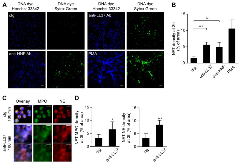
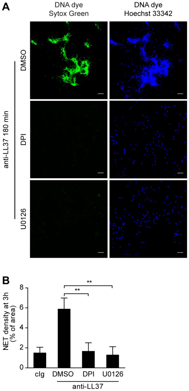
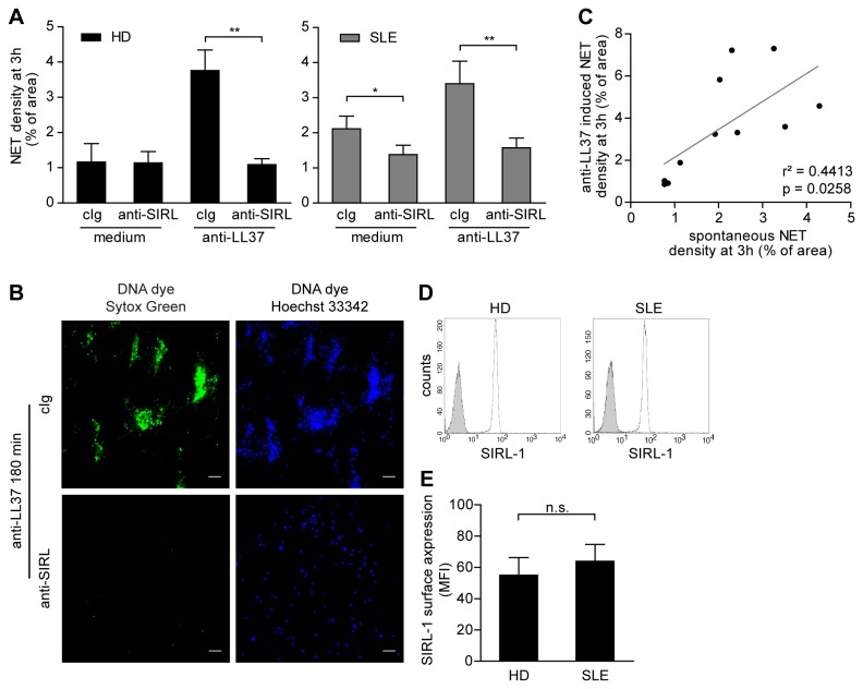
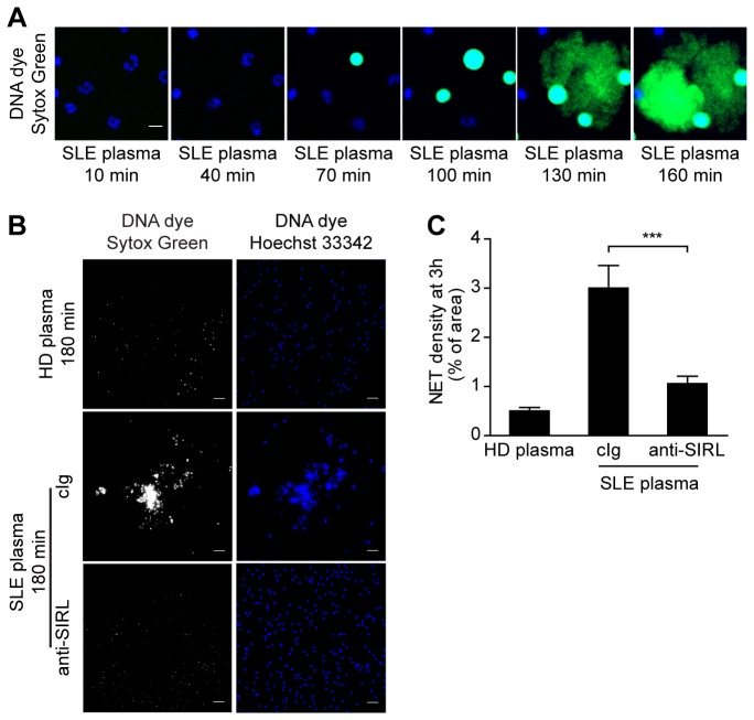
Neutrophil extracellular traps (NETs) have been implicated in the pathogenesis of systemic Lupus erythematosus (SLE), since netting neutrophils release potentially immunogenic autoantigens including histones, LL37, human neutrophil peptide (HNP), and self-DNA. In turn, these NETs activate plasmacytoid dendritic cells resulting in aggravation of inflammation and disease. How suppression of NET formation can be targeted for treatment has not been reported yet. Signal Inhibitory Receptor on Leukocytes-1 (SIRL-1) is a surface molecule exclusively expressed on phagocytes. We recently identified SIRL-1 as a negative regulator of human neutrophil function. Here, we determine whether ligation of SIRL-1 prevents the pathogenic release of NETs in SLE. Peripheral blood neutrophils from SLE patients with mild to moderate disease activity and healthy donors were freshly isolated. NET release was assessed spontaneously or after exposure to anti-neutrophil antibodies or plasma obtained from SLE patients. The formation of NETs was determined by microscopic evaluation using DNA dyes and immunostaining of NET components, as well as by live cell imaging. We show that SLE neutrophils spontaneously release NETs. NET formation is enhanced by stimulation with antibodies against LL37. Inhibition of nicotinamide adenine dinucleotide phosphate (NADPH) oxidase activity and MEK-ERK signaling prevents NET release in response to these antibodies. Signaling via the inhibitory receptor SIRL-1 was induced by ligation with anti-SIRL-1 specific antibodies. Both spontaneous and anti-neutrophil antibody-induced NET formation is suppressed by engagement of SIRL-1. Furthermore, NET release by healthy neutrophils exposed to SLE plasma is inhibited by SIRL-1 ligation. Thus, SIRL-1 engagement can dampen spontaneous and anti-neutrophil antibody-induced NET formation in SLE, likely by suppressing NAPDH oxidase and MEK-ERK activity. Together, these findings reveal a regulatory role for SIRL-1 in NET formation, potentially providing a novel therapeutic target to break the pathogenic loop in SLE.
Conflict of interest statement
Figures
Similar articles
-
Signal inhibitory receptor on leukocytes-1 regulates the formation of the neutrophil extracellular trap in rheumatoid arthritis.Mol Immunol. 2022 Nov;151:242-251. doi: 10.1016/j.molimm.2022.09.008. Epub 2022 Sep 28. Mol Immunol. 2022. PMID: 36182788
-
Neutrophil Extracellular Trap Mitochondrial DNA and Its Autoantibody in Systemic Lupus Erythematosus and a Proof-of-Concept Trial of Metformin.Arthritis Rheumatol. 2015 Dec;67(12):3190-200. doi: 10.1002/art.39296. Arthritis Rheumatol. 2015. PMID: 26245802 Clinical Trial.
-
Caught in a Trap? Proteomic Analysis of Neutrophil Extracellular Traps in Rheumatoid Arthritis and Systemic Lupus Erythematosus.Front Immunol. 2019 Mar 11;10:423. doi: 10.3389/fimmu.2019.00423. eCollection 2019. Front Immunol. 2019. PMID: 30915077 Free PMC article.
-
Neutrophils in the Pathogenesis of Rheumatoid Arthritis and Systemic Lupus Erythematosus: Same Foe Different M.O.Front Immunol. 2021 Mar 4;12:649693. doi: 10.3389/fimmu.2021.649693. eCollection 2021. Front Immunol. 2021. PMID: 33746988 Free PMC article. Review.
-
At the Bedside: Neutrophil extracellular traps (NETs) as targets for biomarkers and therapies in autoimmune diseases.J Leukoc Biol. 2016 Feb;99(2):265-78. doi: 10.1189/jlb.5BT0615-234R. Epub 2015 Dec 11. J Leukoc Biol. 2016. PMID: 26658004 Free PMC article. Review.
Cited by
-
Role of negative regulation of immune signaling pathways in neutrophil function.J Leukoc Biol. 2017 Dec 19:10.1002/JLB.3MIR0917-374R. doi: 10.1002/JLB.3MIR0917-374R. Online ahead of print. J Leukoc Biol. 2017. PMID: 29345376 Free PMC article. Review.
-
VSTM-v1, a potential myeloid differentiation antigen that is downregulated in bone marrow cells from myeloid leukemia patients.J Hematol Oncol. 2015 Mar 15;8:25. doi: 10.1186/s13045-015-0118-4. J Hematol Oncol. 2015. PMID: 25887911 Free PMC article.
-
Innate Immune Dysregulation in the Development of Cardiovascular Disease in Lupus.Curr Rheumatol Rep. 2019 Jul 23;21(9):46. doi: 10.1007/s11926-019-0842-9. Curr Rheumatol Rep. 2019. PMID: 31338604 Review.
-
The Role of Immune Checkpoint Receptors in Regulating Immune Reactivity in Lupus.Cells. 2019 Oct 8;8(10):1213. doi: 10.3390/cells8101213. Cells. 2019. PMID: 31597242 Free PMC article. Review.
-
Immune inhibitory receptor agonist therapeutics.Front Immunol. 2025 Mar 26;16:1566869. doi: 10.3389/fimmu.2025.1566869. eCollection 2025. Front Immunol. 2025. PMID: 40207220 Free PMC article. Review.
References
-
- Villanueva E, Yalavarthi S, Berthier CC, Hodgin JB, Khandpur R et al. (2011) Netting neutrophils induce endothelial damage, infiltrate tissues, and expose immunostimulatory molecules in systemic lupus erythematosus. J Immunol 187: 538-552. doi:10.4049/jimmunol.1100450. PubMed: 21613614. - DOI - PMC - PubMed
Publication types
MeSH terms
Substances
LinkOut - more resources
Full Text Sources
Other Literature Sources
Medical
Molecular Biology Databases
Miscellaneous
