. 2013 Oct 24;8(10):e79077.
doi: 10.1371/journal.pone.0079077. eCollection 2013.

Acute physiological stress promotes clustering of synaptic markers and alters spine morphology in the hippocampus

Affiliations

Acute physiological stress promotes clustering of synaptic markers and alters spine morphology in the hippocampus

Veronica Sebastian et al. PLoS One. .

Abstract

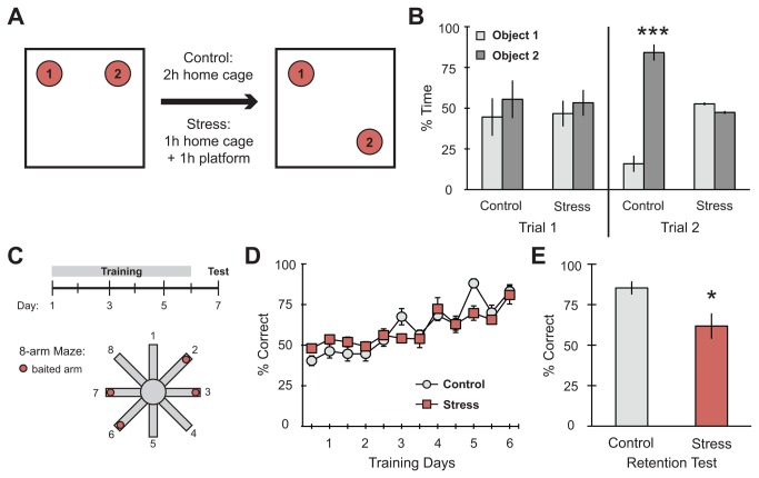
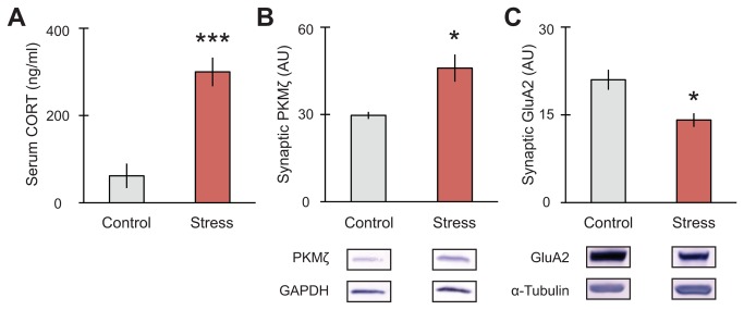
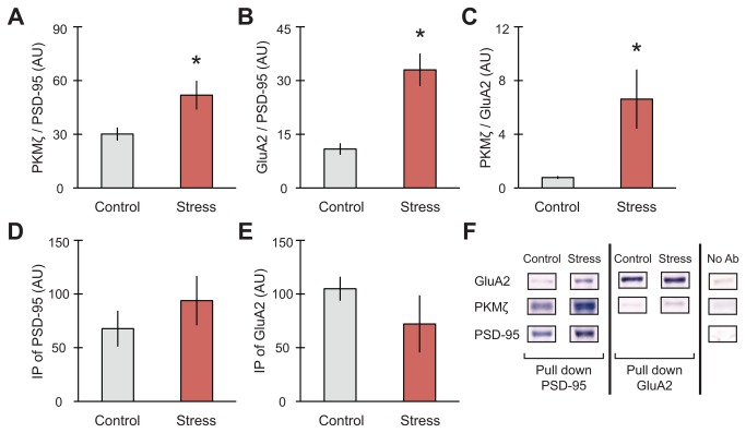
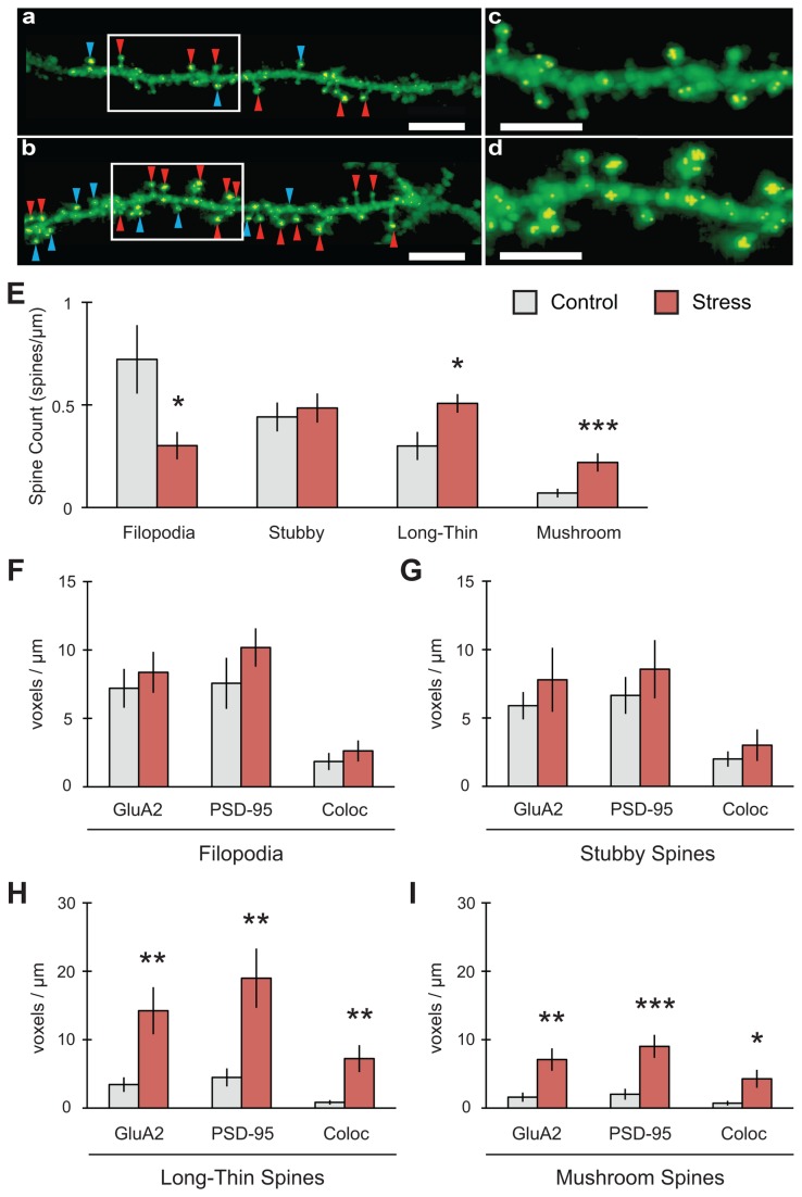
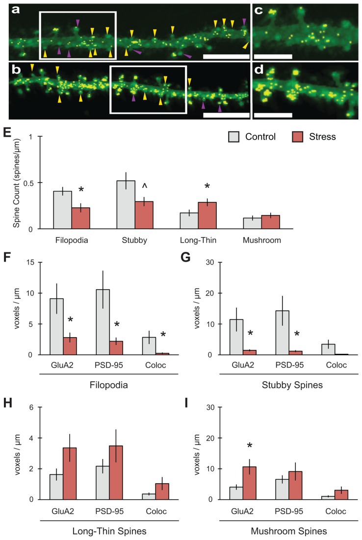
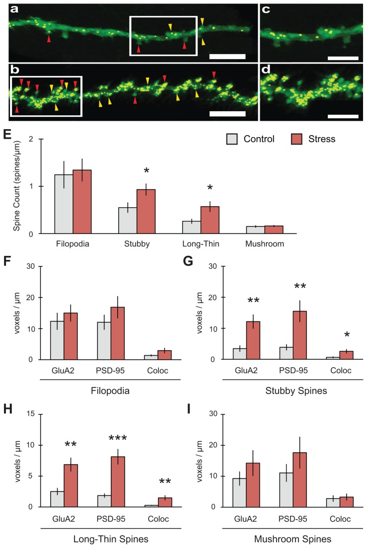
GluA2-containing AMPA receptors and their association with protein kinase M zeta (PKMζ) and post-synaptic density-95 (PSD-95) are important for learning, memory and synaptic plasticity processes. Here we investigated these synaptic markers in the context of an acute 1h platform stress, which can disrupt spatial memory retrieval for a short-term memory on the object placement task and long-term memory retrieval on a well-learned radial arm maze task. Acute stress increased serum corticosterone and elevated the expression of synaptic PKMζ while decreasing synaptic GluA2. Using co-immunoprecipitation, we found that this stressor promotes the clustering of GluA2, PKMζ and PSD-95, which is consistent with effects reported from overexpression of PKMζ in cell culture. Because PKMζ overexpression has also been shown to induce spine maturation in culture, we examined how stress impacts synaptic markers within changing spines across various hippocampal subfields. To achieve this, we employed a new technique combining Golgi staining and immmunohistochemistry to perform 3D reconstruction of tertiary dendrites, which can be analyzed for differences in spine types and the colocalization of synaptic markers within these spines. In CA1, stress increased the densities of long-thin and mushroom spines and the colocalization of GluA2/PSD-95 within these spines. Conversely, in CA3, stress decreased the densities of filopodia and stubby spines, with a concomitant reduction in the colocalization of GluA2/PSD-95 within these spines. In the outer molecular layer (OML) of the dentate gyrus (DG), stress increased both stubby and long-thin spines, together with greater GluA2/PSD-95 colocalization. These data reflect the rapid effects of stress on inducing morphological changes within specific hippocampal subfields, highlighting a potential mechanism by which stress can modulate memory consolidation and retrieval.


Conflict of interest statement

Competing Interests: The authors have declared that no competing interests exist.

Figures

Figure 1
Figure 1. Spatial memory on the object placement and radial arm maze tasks is impaired after platform stress.
(A) Schematic diagram of the object placement task experimental design. (B) There were no significant differences in time spent exploring objects during Trial 1 of the object placement task prior to stress. Controls showed a significant increase in exploration of the object in the novel location (object 2) while stress subjects failed to make a dissociation between objects (n = 6 control, 7 stress). (C) Schematic diagram of the radial arm maze task experimental design. (D) Rats learned the radial arm maze equivalently and significantly improved their performance over training days prior to stress (n = 5 control, 5 stress). (E) Stress prior to the retention test impaired memory retrieval 24h after the last training trial. For all graphs, *p<0.05, ***p<0.001.
Figure 2
Figure 2. Stress increases serum corticosterone and differentially affects synaptic markers for memory in the hippocampus.
(A) Serum corticosterone increased immediately after 1h elevated platform stress (n = 6 control, 10 stress). (B) Synaptic PKMζ in hippocampus increased with platform stress while (C) synaptic GluA2 expression decreased after stress (n = 4 control, 8 stress). (D) Representative blots shown. For all graphs, *p<0.05, ***p<0.001.
Figure 3
Figure 3. Acute stress increases synaptic clustering of GluA2, PKMζ and PSD-95 in hippocampus.
(A) Co-IP of PKMζ with PSD-95 significantly increased (n = 6 control, 8 stress), as did (B) Co-IP of PSD-95 with GluA2 (n = 5 control, 8 stress) and (C) Co-IP of PKMζ with GluA2 (n = 6 control, 6 stress). Overall levels of PSD-95 (D) or GluA2 (E) did not differ between conditions. (F) Representative immunoblots for IP shown. For all graphs, *p<0.05.
Figure 4
Figure 4. Stress increases both mature spines and colocalization of GluA2 with PSD-95 in area CA1.
(A-D) Representative 2D reconstruction of dendrites for control (A, C) and stress (B, D) conditions (scale bar = 5mm for A-B; 3mm for C-D). Golgi-Cox indicated in green, colocalization of synaptic markers in yellow. Red arrowheads indicate long-thin spines, blue arrowheads indicate mushroom spines. (E) Stress increased long-thin (n = 10 control dendrites, 12 stress) and mushroom (n = 11 control, 12 stress) spine counts with a concomitant decrease in filopodia (n = 10 control, 11 stress) and no change in stubby spines (n = 11 control, 12 stress). (F-G) No changes in GluA2, PSD-95 or their colocalization were found in either filopodia (n = 10 control, 8 stress) or stubby spines (n = 12 control, 12 stress). (H) Long-thin spines showed increases in GluA2, PSD-95 and their colocalization (n = 10 control, 10 stress). (I) Mushroom spines showed increases in GluA2, PSD-95 and in their colocalization (n = 11 control, 9 stress). For all graphs, *p<0.05, **p<0.01, ***p<0.001.
Figure 5
Figure 5. Stress reduces both immature spines and colocalization of GluA2 with PSD-95 in area CA3.
(A-D) Representative 2D reconstruction of dendrites for control (A, C) and stress (B, D) conditions (scale bar = 5mm for A-B; 3mm for C-D). Golgi-Cox indicated in green, colocalization of synaptic markers in yellow. Yellow arrowheads indicate stubby spines, purple arrowheads indicate filopodia. (E) Stress decreased filopodia (n = 11 control dendrites, 9 stress) with a concomitant increase in long-thin spines (n = 11 control, 11 stress). Stubby spines (n = 12 control, 11 stress) also demonstrated a trend towards decreased expression, while mushroom spines (n = 12 control, 11 stress) showed no change overall. (F) Filopodia showed a decrease in GluA2, PSD-95 and in their colocalization (n = 9 control, 8 stress). (G) Stubby spines showed a decrease in GluA2 and PSD-95 but no significant change in their colocalization (n = 9 control, 8 stress). (H) No changes in GluA2, PSD-95 or their colocalization were found in long-thin spines (n = 11 control, 11 stress). (I) Stress increased GluA2 expression in mushroom spines but had no effect on PSD-95 or colocalization (n = 12 control, 9 stress). For all graphs, ^p=0.05, *p<0.05.
Figure 6
Figure 6. Stress selectively increases both mature and immature spine types along with colocalization of GluA2 with PSD-95 in the outer molecular layer of the dentate gyrus.
(A-D) Representative 2D reconstruction of dendrites for control (A, C) and stress (B, D) conditions (scale bar = 5mm for A-B; 3mm for C-D). Golgi-Cox indicated in green, colocalization of synaptic markers in yellow. Yellow arrowheads indicate stubby spines, red arrowheads indicate long-thin spines. (E) Stress increased stubby (n = 11 control dendrites, 11 stress) and long-thin (n = 12 control, 10 stress) spine counts. (F) No changes in GluA2, PSD-95 or their colocalization were observed in filopodia (n = 11 control, 10 stress). (G) Stubby spines showed increases in GluA2, PSD-95 and their colocalization (n = 10 control, 10 stress). (H) Long-thin spines showed increases in GluA2, PSD-95 and their colocalization (n = 12 control, 12 stress). (I) No changes were observed in mushroom spines (n = 12 control, 12 stress). For all graphs, *p<0.05, **p<0.01, ***p<0.001.

References

    1. Joëls M, Fernandez G, Roozendaal B (2011) Stress and emotional memory: a matter of timing. Trends Cogn Sci 15: 280-288. doi:10.1016/j.tics.2011.04.004. PubMed: 21571575. - DOI - PubMed
    1. Leuner B, Shors TJ (2012) Stress, anxiety, and dendritic spines: What are the connections? Neuroscience, 251: 108–19. doi:10.1016/j.neuroscience.2012.04.021. PubMed: 22522470. - DOI - PubMed
    1. Joëls M, Baram TZ (2009) The neuro-symphony of stress. Nat Rev Neurosci 10: 459-466. PubMed: 19339973. - PMC - PubMed
    1. Maras PM, Baram TZ (2012) Sculpting the hippocampus from within: stress, spines, and CRH. Trends Neurosci 35: 315-324. doi:10.1016/j.tins.2012.01.005. PubMed: 22386641. - DOI - PMC - PubMed
    1. Gourley SL, Kedves AT, Olausson P, Taylor JR (2009) A history of corticosterone exposure regulates fear extinction and cortical NR2B, GluR2/3, and BDNF. Neuropsychopharmacology 34: 707-716. doi:10.1038/npp.2008.123. PubMed: 18719621. - DOI - PMC - PubMed

Publication types

LinkOut - more resources