. 2013 Oct 24;7(10):e2518.
doi: 10.1371/journal.pntd.0002518. eCollection 2013.

Suppression of E. multilocularis hydatid cysts after ionizing radiation exposure

Affiliations

Suppression of E. multilocularis hydatid cysts after ionizing radiation exposure

Xin Zhou et al. PLoS Negl Trop Dis. .

Abstract

Background: Heavy-ion therapy has an advantage over conventional radiotherapy due to its superb biological effectiveness and dose conformity in cancer therapy. It could be a potential alternate approach for hydatid cyst treatment. However, there is no information currently available on the cellular and molecular basis for heavy-ion irradiation induced cell death in cystic echinococcosis.

Methododology/principal findings: LD50 was scored by protoscolex death. Cellular and ultrastructural changes within the parasite were studied by light and electron microscopy, mitochondrial DNA (mtDNA) damage and copy number were measured by QPCR, and apoptosis was determined by caspase 3 expression and caspase 3 activity. Ionizing radiation induced sparse cytoplasm, disorganized and clumped organelles, large vacuoles and devoid of villi. The initial mtDNA damage caused by ionizing radiation increased in a dose-dependent manner. The kinetic of DNA repair was slower after carbon-ion radiation than that after X-rays radiation. High dose carbon-ion radiation caused irreversible mtDNA degradation. Cysts apoptosis was pronounced after radiation. Carbon-ion radiation was more effective to suppress hydatid cysts than X-rays.

Conclusions: These studies provide a framework to the evaluation of attenuation effect of heavy-ion radiation on cystic echinococcosis in vitro. Carbon-ion radiation is more effective to suppress E. multilocularis than X-rays.


Conflict of interest statement

The authors have declared that no competing interests exist.

Figures

Figure 1
Figure 1. Percent lethality versus radiation dose.
The data are fitted to a sigmoidal dose response function: y = a+b/(1+exp(-(x(-c)/d)), where y is the percent lethality, x is the dose in Gy, respectively.
Figure 2
Figure 2. DNA damage repair and mtDNA copy number in irradiated hydatid cysts.
(A) Quantification of mtDNA damage by long-PCR amplification of total DNA isolated from E. multilocularis hydatid cysts after 5–30 Gy X-rays or carbon-ion radiation. MtDNA damage was indicated by reduced PCR amplification. (B) Repair kinetics of mtDNA in E. multilocularis hydatid cysts within 72 hours after 30 Gy X-rays or carbon-ion radiation. (C) Quantification of mtDNA copy number by real-time PCR. Error bars represent the SD, each done in at least triplicate. *Statistical significant at p<0.01.
Figure 3
Figure 3. Representative thin-section electron microscopy of E. multilocularis hydatid cysts from control and irradiated cultures.
Examples of control (A and B), 30 Gy X-rays irradiated (C and D) and 30 Gy carbon-ion irradiated (E and F) parasites are shown. A and B, the majority of the parasites displayed organized internal structures and their organelles were intact and clear. C—F, in irradiated samples, parasites displayed abnormal morphologies that included clumped organelles, abnormally large vacuoles, and generally disorganized structure. 1: germinal layer; 2: laminated layer; 3: border between germinal layer and laminated layer; 4: mitochondria; 5: vacuole; 6: damaged organelle.
Figure 4
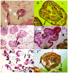
Figure 4. Representative light microscopy of E. multilocularis hydatid cysts from control and irradiated cultures.
Examples of control (A and B), 30 Gy X-rays irradiated(C and D) and 30 Gy carbon-ion irradiated (E and F) parasites are shown. A, The protoscolex, germinal layer and cuticles were intact and clear (×40) in unirradiated samples. B. Protoscolex was visible and intact (×100). C and D, after 30 Gy X-rays irradiation, parasites displayed abnormal morphologies that included protoscolex eversion (1) and detachment of germinal layer from cuticles (2). E, After 30 Gy carbon-ion irradiation, loss of suction cups and scolex hooks, protoscolex contraction (3) were extensive in parasites.
Figure 5
Figure 5. Apoptosis in 30 Gy X-rays and carbon-ion irradiated E. multilocularis hydatid cysts.
A, Caspase 3 expression in hydatid cysts with and without 30 Gy ionizing radiation. B, Caspase 3 like activity was measured as the difference in pNA production between the samples with and without ionizing radiation. Error bars represent the SD, each done in at least triplicate. *Statistical significant at p<0.01.

References

    1. Eckert J, Deplazes P (2004) Biological, epidemiological, and clinical aspects of echinococcosis, a zoonosis of increasing concern. Clin Microbiol Rev 17: 107–135. - PMC - PubMed
    1. Bao YX, Zhang YF, Ni YQ, Xie ZR, Qi HZ, et al. (2011) [Effect of 6 MeV radiotherapy on secondary Echinococcus multilocularis infection in rats]. Zhongguo Ji Sheng Chong Xue Yu Ji Sheng Chong Bing Za Zhi 29: 127–129. - PubMed
    1. Bao YX, Mao R, Qi HZ, Zhang YF, Ni YQ, et al. (2011) [X-ray irradiation against Echinococcus multilocularis protoscoleces in vitro]. Zhongguo Ji Sheng Chong Xue Yu Ji Sheng Chong Bing Za Zhi 29: 208–211. - PubMed
    1. Ulger S, Barut H, Tunc M, Aydin E, Aydinkarahaliloglu E, et al. (2013) Radiation therapy for resistant sternal hydatid disease. Strahlenther Onkol 189: 508–9. - PubMed
    1. Phillips R, Murikami K (1960) Preliminary neoplasms of the liver. Results of radiation therapy. Cancer 13: 714–720. - PubMed

Publication types