Case Reports
doi: 10.4103/2231-0746.119222.
Congenital muscular torticollis
Affiliations
- PMID: 24205484
- PMCID: PMC3814673
- DOI: 10.4103/2231-0746.119222
Item in Clipboard
Case Reports
Congenital muscular torticollis
Ann Maxillofac Surg.
2013 Jul.
Abstract
Congenital muscular torticollis (CMT) is a rare congenital musculoskeletal disorder characterized by unilateral shortening of the sternocleidomastoid muscle (SCM). It presents in newborn infants or young children with reported incidence ranging from 0.3% to 2%. Owing to effective shortening of SCM on the involved side there is ipsilateral head tilt and contralateral rotation of the face and chin. This article reports a case of CMT in a 3½-year-old male child successfully managed by surgical release of the involved SCM followed by physiotherapy.
Keywords: Congenital; sternocleidomastoid muscle; tenotomy; torticollis.
Conflict of interest statement
Figures
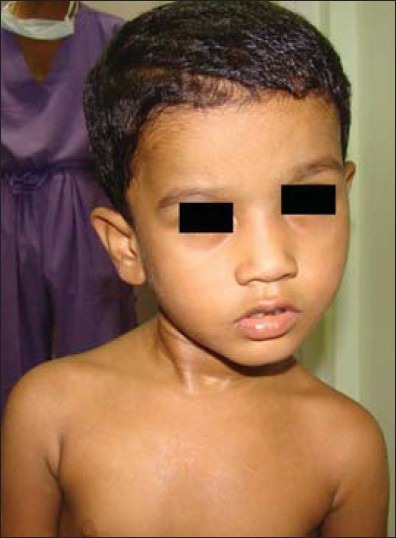
Preoperative clinical photograph showing the child with congenital muscular torticollis affecting the right sternocleidomastoid muscle
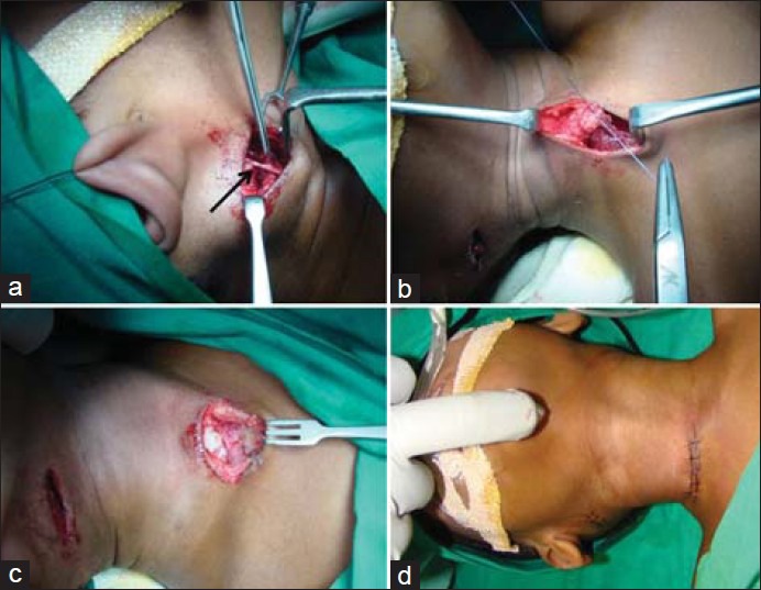
Intraoperative photographs. (a) Incision place over mastoid region, the overlying greater auricular nerve (arrow) was identified and protected during dissection. (b) Bot h the clavicular and sternal heads of the sternocleidomastoid muscle were identified and then divided. Sternal end was then sutured to the clavicular cut end in an oblique line to achieve muscle lengthening. (c) The excised lower end of SCM. (d) Closure done and complete neck extension was achieved without any strain intraoperatively
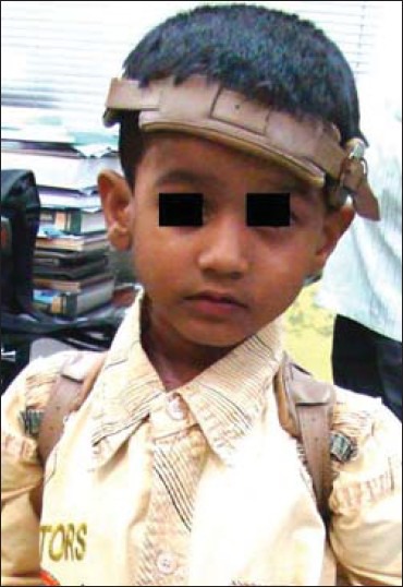
Torticollis brace worn by the patient postoperatively
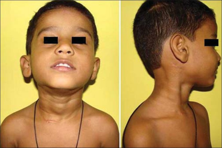
At 12 months postoperative photographs showing straight neck with a normal range of neck movement
References
-
- Tubby AH. 2nd ed. Vol. 1. London, England: MacMillan; 1912. Deformities and Diseases of Bones and Joints; p. 56.
-
- Wei JL, Schwartz KM, Weaver AL, Orvidas LJ. Pseudotumor of infancy and congenital muscular torticollis: 170 cases. Laryngoscope. 2001;111:688–95. - PubMed
-
- Davids JR, Wenger DR, Mubarak SJ. Congenital muscular torticollis: Sequela of intrauterine or perinatal compartment syndrome. J Pediatr Orthop. 1993;13:141–7. - PubMed
-
- Tang S, Liu Z, Quan X, Qin J, Zhang D. Sternocleidomastoid pseudotumor of infants and congenital muscular torticollis: Fine-structure research. J Pediatr Orthop. 1998;18:214–8. - PubMed
-
- Hollier L, Kim J, Grayson BH, McCarthy JG. Congenital muscular torticollis and the associated craniofacial changes. Plast Reconstr Surg. 2000;105:827–35. - PubMed
Publication types
LinkOut - more resources
Full Text Sources
Other Literature Sources