

Endoscopically visualized lesions, histologic findings, and bacterial invasion in the gastrointestinal mucosa of dogs with acute hemorrhagic diarrhea syndrome
- PMID: 24205886
- PMCID: PMC4895553
- DOI: 10.1111/jvim.12236
Endoscopically visualized lesions, histologic findings, and bacterial invasion in the gastrointestinal mucosa of dogs with acute hemorrhagic diarrhea syndrome
Abstract
Background: Etiology of hemorrhagic gastroenteritis (HGE) syndrome in dogs is unknown and histopathologic and microbial investigations have only been performed post mortem.
Objective: To identify characteristic intra vitam endoscopic and histologic mucosal lesions, as well as bacterial species, within the mucosa of dogs with HGE.
Animals: Ten dogs diagnosed with HGE were included. Eleven dogs with gastroduodenoscopy and different intestinal diseases were used as controls for microbial changes. Dogs pretreated with antibiotics or diagnosed with any disease known to cause bloody diarrhea were excluded from the study.
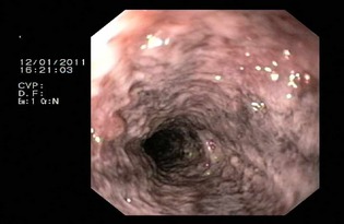
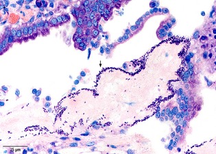
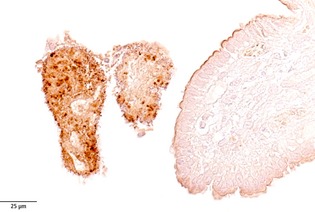
Methods: In this prospective study, gastrointestinal biopsies were collected from 10 dogs with HGE. Endoscopic and histologic changes were assessed according to WSAVA guidelines. Biopsies from the stomach, duodenum, ileum, and colon were investigated by histology and by immunohistochemistry for the presence of Clostridium spp. and parvovirus. The first duodenal biopsy taken with a sterile forceps was submitted for bacterial culture.
Results: Acute mucosal lesions were only found in the intestines, not in the stomach. Clostridium spp., identified as Clostridium perfringens in 6/9 cases, were detected on the small intestinal mucosa in all dogs with HGE, either by culture or immunohistopathology. In the control group, C. perfringens could only be cultured in one of 11 dogs.
Conclusions and clinical importance: The results of this study demonstrate an apparent association between C. perfringens and the occurrence of acute hemorrhagic diarrhea. The term "HGE," which implies the involvement of the stomach, should be renamed as "acute hemorrhagic diarrhea syndrome."
Keywords: Acute emorrhagic diarrhea syndrome; Bloody diarrhea; Clostridium perfringens; Hemorrhagic gastroenteritis.
Copyright © 2013 by the American College of Veterinary Internal Medicine.
Figures
References
-
- Burrows C. Canine Hemorrhagic gastroenteritis. J Am Anim Hosp Assoc 1977;13:451–458.
-
- Unterer S, Strohmeyer K, Kruse BD, et al. Treatment of aseptic dogs with hemorrhagic gastroenteritis with amoxicillin/clavulanic acid: A prospective blinded study. J Vet Intern Med 2011;25:973–979. - PubMed
-
- Spielman B, Garvey M. Hemorrhagic gastroenteritis in 15 Dogs. J Am Anim Hosp Assoc 1993;29:341–344.
-
- Holt PE. Haemorrhagic gastroenteritis in a dog. Vet Rec 1979;104:150. - PubMed
-
- Cave NJ, Marks SL, Kass PH, et al. Evaluation of a routine diagnostic fecal panel for dogs with diarrhea. J Am Vet Med Assoc 2002;221:52–59. - PubMed
MeSH terms
LinkOut - more resources
Full Text Sources
Other Literature Sources
Medical
Miscellaneous
