Review
. 2014 Jun 10;20(17):2815-37.
doi: 10.1089/ars.2013.5703. Epub 2014 Jan 16.

New insights on NOX enzymes in the central nervous system

Affiliations
Review

New insights on NOX enzymes in the central nervous system

Zeynab Nayernia et al. Antioxid Redox Signal. .

Abstract

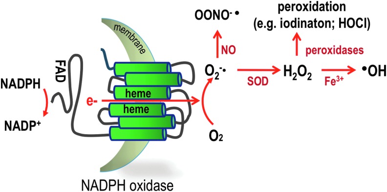
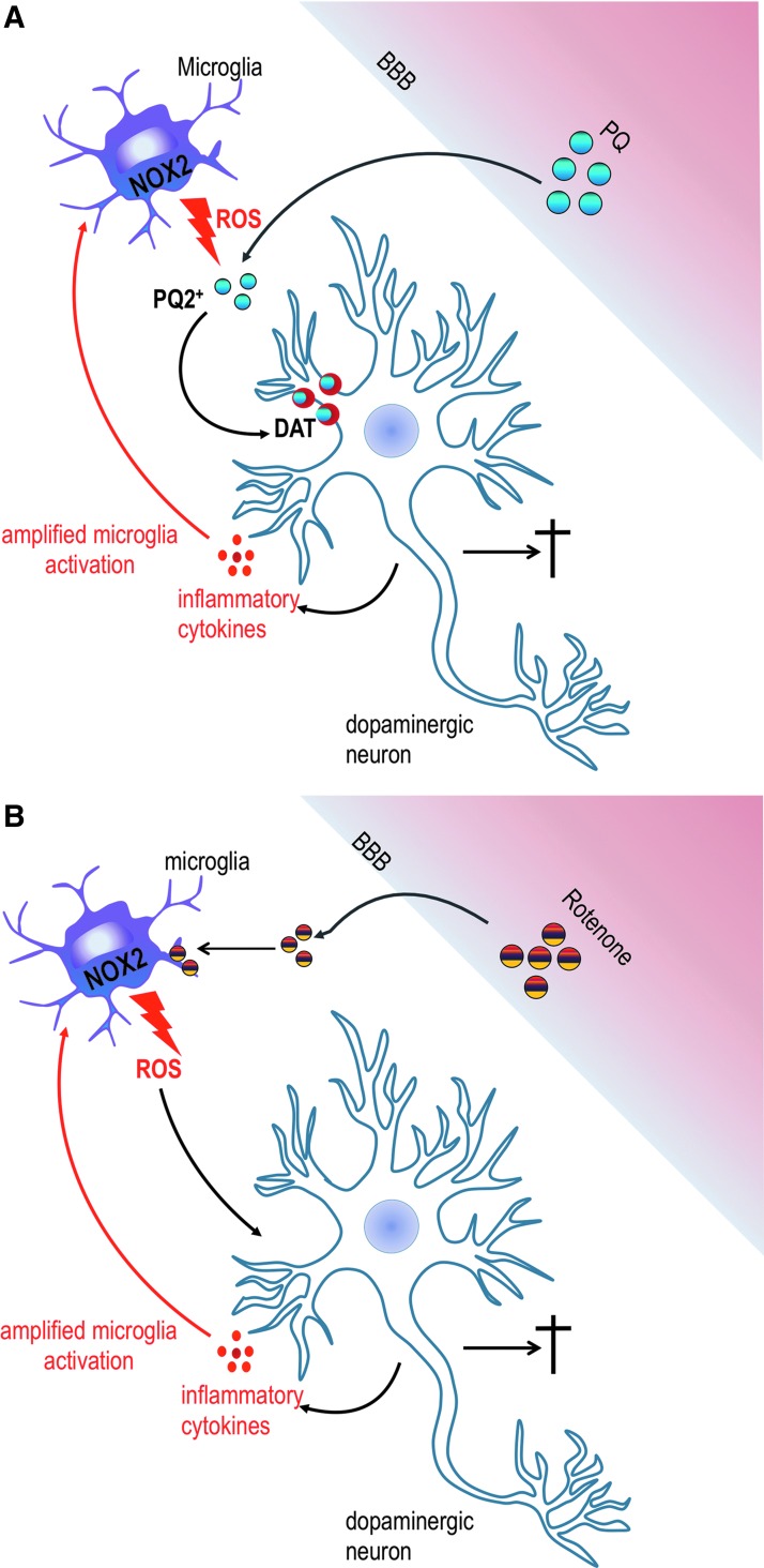
Significance: There is increasing evidence that the generation of reactive oxygen species (ROS) in the central nervous system (CNS) involves the NOX family of nicotinamide adenine dinucleotide phosphate oxidases. Controlled ROS generation appears necessary for optimal functioning of the CNS through fine-tuning of redox-sensitive signaling pathways, while overshooting ROS generation will lead to oxidative stress and CNS disease.

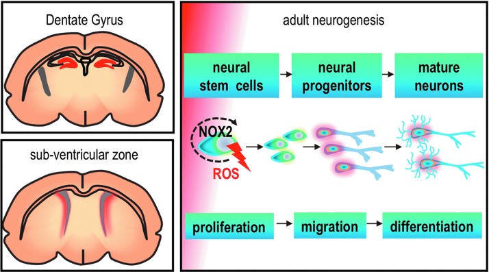
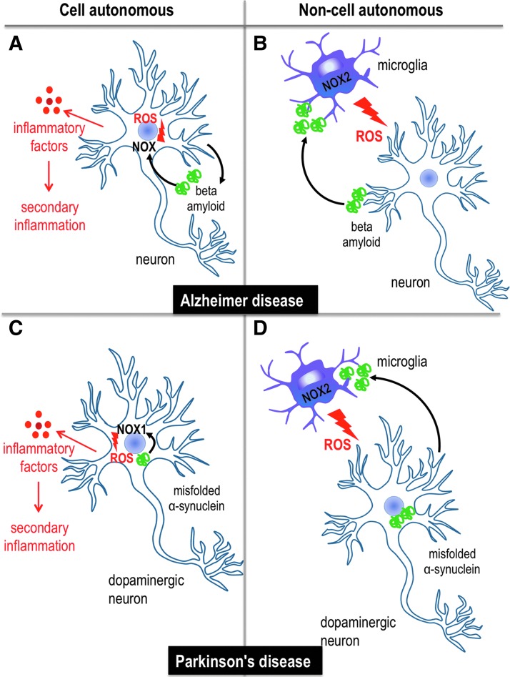
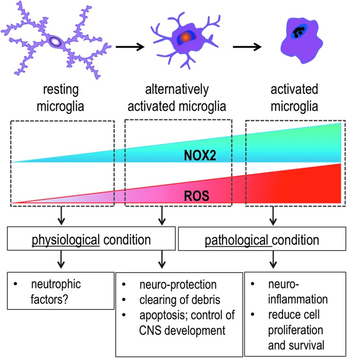
Recent advances: NOX enzymes are not only restricted to microglia (i.e. brain phagocytes) but also expressed in neurons, astrocytes, and the neurovascular system. NOX enzymes are involved in CNS development, neural stem cell biology, and the function of mature neurons. While NOX2 appears to be a major source of pathological oxidative stress in the CNS, other NOX isoforms might also be of importance, for example, NOX4 in stroke. Globally speaking, there is now convincing evidence for a role of NOX enzymes in various neurodegenerative diseases, cerebrovascular diseases, and psychosis-related disorders.

Critical issues: The relative importance of specific ROS sources (e.g., NOX enzymes vs. mitochondria; NOX2 vs. NOX4) in different pathological processes needs further investigation. The absence of specific inhibitors limits the possibility to investigate specific therapeutic strategies. The uncritical use of non-specific inhibitors (e.g., apocynin, diphenylene iodonium) and poorly validated antibodies may lead to misleading conclusions.

Future directions: Physiological and pathophysiological studies with cell-type-specific knock-out mice will be necessary to delineate the precise functions of NOX enzymes and their implications in pathomechanisms. The development of CNS-permeant, specific NOX inhibitors will be necessary to advance toward therapeutic applications.


Figures

<b>FIG. 1.</b>
FIG. 1.
Schematic representation of NOX NADPH oxidases. NOX enzymes are transmembrane proteins that catalyze the transfer of two electrons through biological membranes using intracellular NADPH as an electron donor and extracellular molecular oxygen as an acceptor. The primary product of this reaction is the superoxide anion O2−•. From this primary product and depending on the systems, numerous other ROS can be generated. In the presence of NO, O2−• rapidly reacts and forms the highly toxic peroxynitrite or it can be dismutated either by a superoxide dismutase or spontaneously to form hydrogen peroxide, which displays numerous actions as a second messenger and reacts in the presence of peroxidase to form the microbicidal hypochlorous acid in the phagosome or in the presence of Fe3+, the highly reactive hydroxyl radical, which is known to modify lipids and proteins. NADPH, nicotinamide adenine dinucleotide phosphate; NO, nitric oxide; NOX, nicotinamide adenine dinucleotide phosphate-dependent oxidase; ROS, reactive oxygen species. To see this illustration in color, the reader is referred to the web version of this article at www.liebertpub.com/ars
<b>FIG. 2.</b>
FIG. 2.
NOX2 controls neurogenesis of adult neural stem cells. Adult neural stem cells reside in two main regions of the brain: dentate gyrus and sub ventricular zone (regions shown with red color). A controlled level of ROS generation by NOX2 at this early stage of adult neurogenesis is involved with the proliferation and survival of neural stem cells.
<b>FIG. 3.</b>
FIG. 3.
Neuroinflammation and oxidative stress in cell autonomous and/or non-cell autonomous models of neurodegenerative diseases. Both mechanisms are described in models of Alzheimer and Parkinson disease. (A) In Alzheimer diseases, amyloid fragments generated through proteolytic cleavage of the APP are released to the extracellular space. This pathological protein expressed in neurons leads to neuronal damage by ROS produced by NOX and release of inflammatory factors, leading to secondary inflammation. (B) Non-cell autonomous mechanisms involve cells other than neurons, in particular the activation of microglia and ROS production generated by NOX2. In Alzheimer disease, the activation of microglia cells and the generation of ROS are considered to be mediated by beta amyloid fragments that are released extracellularly. (C) In Parkinson disease, intracellular protein aggregates might lead to ROS production via NOX1 and cellular stress, which leads to the generation of inflammatory cytokines. (D) In the non-cell autonomous state, misfolded α-synuclein protein fragments are secreted from neurons and may, therefore, activate microglia cells, which leads to ROS generation by NOX2 and oxidative stress. APP, amyloid precursor protein. To see this illustration in color, the reader is referred to the web version of this article at www.liebertpub.com/ars
<b>FIG. 4.</b>
FIG. 4.
The dual function of microglia NOX2 in the CNS: physiology and pathology. At a resting stage, microglia is highly branched, generates little ROS, and shows very low NOX2 expression. An elevated and controlled level of ROS generated by microglial NOX2 is involved in the physiological condition and healthy non-inflammatory state of the CNS, which plays a role in programmed cell death of cells in the brain during CNS development. After CNS insult, microglial NOX2 is induced. Microglia develop into an active state in which they display a protective phenotype (alternative activation) in order to clear debris and support neuronal survival. In some instances, the inflammatory signals such as environmental toxins and fragments of proteins are not removed from the CNS. In response, microglia become amoeboid and enter an over-activated and neurotoxic stage that is characterized by high NOX2 expression and ROS generation. CNS, central nervous system. To see this illustration in color, the reader is referred to the web version of this article at www.liebertpub.com/ars
<b>FIG. 5.</b>
FIG. 5.
Activation of microglia NOX2 via environmental toxics through two unique mechanisms in Parkinson disease. (A) If PQ is oxidized (PQ2+) by ROS generated by NOX2, it becomes a substrate for the DAT, which actively transports it into dopaminergic neurons. Oxidized PQ might be directly neurotoxic and also lead to a release of inflammatory factors by neurons. This eventually leads to amplification of the signal (red arrow) and death of dopaminergic neurons. (B) Rotenone directly interacts with microglia NOX2 and elicits ROS production and damage of dopaminergic neurons. This leads to a release of inflammatory factors, the recruitment of more microglia cells, and, eventually, dopaminergic neuron death. DAT, dopamine transporter; PQ, paraquat. To see this illustration in color, the reader is referred to the web version of this article at www.liebertpub.com/ars
<b>FIG. 6.</b>
FIG. 6.
Hoffer and Osmond hypothesis for oxidative stress-mediated psychosis. Ex vivo oxidation of epinephrine (also known as adrenaline) generates adrenochrome, a potent psychotropic compound that is characterized by its pink color in the solution. This oxidized epinephrine is consumed as a recreational drug and its street name is “pink adrenaline.” A speculative, but interesting hypothesis suggests that increased oxidation in psychotic patients may lead to the generation of adrenochrome, which might contribute to characteristic symptoms of schizophrenia, such as hallucinations and delusion.

References

    1. Aguzzi A, Barres BA, and Bennett ML. Microglia: scapegoat, saboteur, or something else? Science 339: 156–161, 2013 - PMC - PubMed
    1. Ali SS, Young JW, Wallace CK, Gresack J, Jeste DV, Geyer MA, Dugan LL, and Risbrough VB. Initial evidence linking synaptic superoxide production with poor short-term memory in aged mice. Brain Res 1368: 65–70, 2011 - PMC - PubMed
    1. Alliot F, Godin I, and Pessac B. Microglia derive from progenitors, originating from the yolk sac, and which proliferate in the brain. Brain Res Dev Brain Res 117: 145–152, 1999 - PubMed
    1. Altenhofer S, Kleikers PW, Radermacher KA, Scheurer P, Rob Hermans JJ, Schiffers P, Ho H, Wingler K, and Schmidt HH. The NOX toolbox: validating the role of NADPH oxidases in physiology and disease. Cell Mol Life Sci 69: 2327–2343, 2012 - PMC - PubMed
    1. Andreu CI, Woehlbier U, Torres M, and Hetz C. Protein disulfide isomerases in neurodegeneration: from disease mechanisms to biomedical applications. FEBS Lett 586: 2826–2834, 2012 - PubMed

Publication types

MeSH terms