Meta-Analysis
. 2013 Nov 10:13:538.
doi: 10.1186/1471-2407-13-538.

The potential of hypoxia markers as target for breast molecular imaging--a systematic review and meta-analysis of human marker expression

Affiliations
Meta-Analysis

The potential of hypoxia markers as target for breast molecular imaging--a systematic review and meta-analysis of human marker expression

Arthur Adams et al. BMC Cancer. .

Abstract

Background: Molecular imaging of breast cancer is a promising emerging technology, potentially able to improve clinical care. Valid imaging targets for molecular imaging tracer development are membrane-bound hypoxia-related proteins, expressed when tumor growth outpaces neo-angiogenesis. We performed a systematic literature review and meta-analysis of such hypoxia marker expression rates in human breast cancer to evaluate their potential as clinically relevant molecular imaging targets.

Methods: We searched MEDLINE and EMBASE for articles describing membrane-bound proteins that are related to hypoxia inducible factor 1α (HIF-1α), the key regulator of the hypoxia response. We extracted expression rates of carbonic anhydrase-IX (CAIX), glucose transporter-1 (GLUT1), C-X-C chemokine receptor type-4 (CXCR4), or insulin-like growth factor-1 receptor (IGF1R) in human breast disease, evaluated by immunohistochemistry. We pooled study results using random-effects models and applied meta-regression to identify associations with clinicopathological variables.

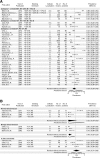
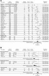
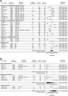
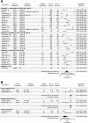
Results: Of 1,705 identified articles, 117 matched our selection criteria, totaling 30,216 immunohistochemistry results. We found substantial between-study variability in expression rates. Invasive cancer showed pooled expression rates of 35% for CAIX (95% confidence interval (CI): 26-46%), 51% for GLUT1 (CI: 40-61%), 46% for CXCR4 (CI: 33-59%), and 46% for IGF1R (CI: 35-70%). Expression rates increased with tumor grade for GLUT1, CAIX, and CXCR4 (all p < 0.001), but decreased for IGF1R (p < 0.001). GLUT1 showed the highest expression rate in grade III cancers with 58% (45-69%). CXCR4 showed the highest expression rate in small T1 tumors with 48% (CI: 28-69%), but associations with size were only significant for CAIX (p < 0.001; positive association) and IGF1R (p = 0.047; negative association). Although based on few studies, CAIX, GLUT1, and CXCR4 showed profound lower expression rates in normal breast tissue and benign breast disease (p < 0.001), and high rates in carcinoma in situ. Invasive lobular carcinoma consistently showed lower expression rates (p < 0.001).

Conclusions: Our results support the potential of hypoxia-related markers as breast cancer molecular imaging targets. Although specificity is promising, combining targets would be necessary for optimal sensitivity. These data could help guide the choice of imaging targets for tracer development depending on the envisioned clinical application.


Figures
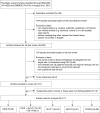
Figure 1
Figure 1
Flowchart for selection of articles describing expression prevalences of the hypoxia markers CAIX, GLUT1, CXCR4, and IGF1R in breast cancer, normal tissue, benign breast disease, and carcinoma in situ , assessed by immunohistochemistry.
Figure 2
Figure 2
Expression prevalence of CAIX. A Systematic literature review of CAIX prevalence in breast cancer assessed by immunohistochemistry, according to reported staining threshold. Legend: Dashed gray reference line: overall random-effects prevalence estimate. Abbreviations: Staining threshold: weak intensity (WI), moderate intensity (MI), strong intensity (SI); Localization: cytoplasm (c), membrane (m); confidence interval (CI); not stated (NS). B Systematic literature review of CAIX prevalence in normal breast tissue, benign breast diseases and carcinoma in situ assessed by immunohistochemistry. Legend: Dashed line represents random effect summary prevalence estimate for invasive cancer within studies reporting also on normal, benign and/or precancerous breast tissue (4 studies). Abbreviations: Staining threshold: weak intensity (WI), moderate intensity (MI), strong intensity (SI); Localization: cytoplasm (c), membrane (m); confidence interval (CI); not stated (NS).
Figure 3
Figure 3
Expression prevalence of GLUT1. A Systematic literature review of GLUT1 prevalence in breast cancer assessed by immunohistochemistry, according to reported staining threshold. Legend: Dashed gray reference line: overall random-effects prevalence estimate. Abbreviations: Staining threshold: weak intensity (WI), moderate intensity (MI), strong intensity (SI); Localization: cytoplasm (c), membrane (m); confidence interval (CI); not stated (NS). B Systematic literature review of GLUT 1 prevalence in normal breast tissue, benign breast diseases and carcinoma in situ assessed by immunohistochemistry. Legend: Dashed line represents random effect summary prevalence estimate for invasive cancer within studies reporting also on normal, benign and/or precancerous breast tissue ( 5 studies). Abbreviations: Staining threshold: weak intensity (WI), moderate intensity (MI), strong intensity (SI); Localization: cytoplasm (c), membrane (m); confidence interval (CI); not stated (NS).
Figure 4
Figure 4
Expression prevalence of CXCR4. A Systematic literature review of CXCR4 prevalence in breast cancer assessed by immunohistochemistry, according to reported staining threshold. Legend: Dashed gray reference line: overall random-effects prevalence estimate. Abbreviations: Staining threshold: weak intensity (WI), moderate intensity (MI), strong intensity (SI); Localization: cytoplasm (c), membrane (m); confidence interval (CI); not stated (NS). B Systematic literature review of CXCR4 prevalence in normal breast tissue, benign breast diseases and carcinoma in situ assessed by immunohistochemistry. Legend: Dashed line represents random effect summary prevalence estimate for invasive cancer within studies reporting also on normal, benign and/or precancerous breast tissue (6 studies). Abbreviations: Staining threshold: weak intensity (WI), moderate intensity (MI), strong intensity (SI); Localization: cytoplasm (c), membrane (m); confidence interval (CI); not stated (NS).
Figure 5
Figure 5
Expression prevalence of IGF1R. A Systematic literature review of IGF1R prevalence in breast cancer assessed by immunohistochemistry, according to reported staining threshold. Legend: Dashed gray reference line: overall random-effects prevalence estimate. Abbreviations: Staining threshold: weak intensity (WI), moderate intensity (MI), strong intensity (SI); Localization: cytoplasm (c), membrane (m); confidence interval (CI); not stated (NS). B Systematic literature review of IGF1R prevalence in normal breast tissue, benign breast diseases and carcinoma in situ assessed by immunohistochemistry. Legend: Dashed line represents random effect summary prevalence estimate for invasive cancer within studies reporting also on normal, benign and/or precancerous breast tissue ( 4 studies). Abbreviations: Staining threshold: weak intensity (WI), moderate intensity (MI), strong intensity (SI); Localization: cytoplasm (c), membrane (m); confidence interval (CI); not stated (NS).

References

    1. Sampath L, Wang W, Sevick-Muraca EM. Near infrared fluorescent optical imaging for nodal staging. J Biomed Opt. 2008;13(4):041312. - PMC - PubMed
    1. Goldenberg DM, Nabi HA. Breast cancer imaging with radiolabeled antibodies. Semin Nucl Med. 1999;13(1):41–48. - PubMed
    1. Mann RM, Hoogeveen YL, Blickman JG, Boetes C. MRI compared to conventional diagnostic work-up in the detection and evaluation of invasive lobular carcinoma of the breast: a review of existing literature. Breast Cancer Res Treat. 2008;13(1):1–14. - PMC - PubMed
    1. Leung JW. New modalities in breast imaging: digital mammography, positron emission tomography, and sestamibi scintimammography. Radiol Clin North Am. 2002;13(3):467–482. - PubMed
    1. Petralia G, Bonello L, Priolo F, Summers P, Bellomi M. Breast MR with special focus on DW-MRI and DCE-MRI. Cancer Imaging. 2011;13:76–90. - PMC - PubMed

Publication types