

Isolated persistent left-sided superior vena cava, giant coronary sinus, atrial tachycardia and heart failure in a child
- PMID: 24206885
- PMCID: PMC3860611
- DOI: 10.1016/j.ihj.2013.08.024
Isolated persistent left-sided superior vena cava, giant coronary sinus, atrial tachycardia and heart failure in a child
Abstract
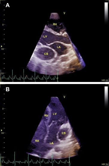
Persistence of a left-sided superior vena cava (PLSVC) with absent right superior vena cava (isolated PLSVC) is a very rare venous malformation and commonly associated with congenital heart disease or alterations of the cardiac situs. We describe an unusual case of a young boy presenting with persistent atrial tachycardia and congestive heart failure. He was detected to have unexplained grossly dilated right atrium, right ventricle with systolic dysfunction and a giant coronary sinus (CS). The dilated CS closely mimicked a pseudo cor-triatriatum on echocardiography. Contrast echocardiography from both arms revealed opacification of the CS before the right atrium. Bilateral upper limb venography confirmed the presence of absent right SVC and isolated persistent left SVC draining into the giant coronary sinus.
Keywords: Atrial tachycardia; Congestive heart failure; Coronary sinus; Persistent LSVC.
Copyright © 2013 Cardiological Society of India. Published by Elsevier B.V. All rights reserved.
Figures


References
-
- Higgs A.G., Paris S., Potter F. Discovery of left-sided superior vena cava during central venous catheterization. Br J Anaesth. 1998;81:260–261. - PubMed
-
- Winter F.S. Persistent left superior vena cava; survey of world literature and report of thirty additional cases. Angiology. 1954;5:90–132. - PubMed
-
- Boussuges A., Ambrosi P., Gainnier M., Quenee V., Saint J.M. Left-sided superior vena cava: diagnosis by magnetic resonance imaging. Intens Care Med. 1997;23:702–770. - PubMed
-
- Pai R.G. Echocardiographic features of persistent left superior vena cava. Echocardiography. 1999;16:435–436. - PubMed
Publication types
MeSH terms
LinkOut - more resources
Full Text Sources
Other Literature Sources
Medical
