Review
. 2013 Nov 7;52(3):291-302.
doi: 10.1016/j.molcel.2013.10.018.

The intertwined roles of transcription and repair proteins

Affiliations
Review

The intertwined roles of transcription and repair proteins

Yick W Fong et al. Mol Cell. .

Abstract

Transcription is apparently risky business. Its intrinsic mutagenic potential must be kept in check by networks of DNA repair factors that monitor the transcription process to repair DNA lesions that could otherwise compromise transcriptional fidelity and genome integrity. Intriguingly, recent studies point to an even more direct function of DNA repair complexes as coactivators of transcription and the unexpected role of "scheduled" DNA damage/repair at gene promoters. Paradoxically, spontaneous DNA double-strand breaks also induce ectopic transcription that is essential for repair. Thus, transcription, DNA damage, and repair may be more physically and functionally intertwined than previously appreciated.


Figures

Figure 1
Figure 1. Major DNA repair pathways in mammals
Exogenous and endogenous genotoxic agents (top) generate a variety of DNA damage, such as single and double strand breaks (SSBs, DSBs), insertions and deletions (indels). Lesions are detected and repaired by four major DNA repair pathways: base excision repair (BER, A), nucleotide excision repair (NER, B), mismatch repair (MMR, C) and recombinational repair (D). Mechanisms of BER, NER and recombinational repair are depicted. For each pathway, factors discussed in this review are in colors. A. Removal of uracil from DNA by BER. Thymine DNA glycosylase (TDG) removes the nitrogenous base and generates an abasic site (*). Apurinic/apyrimidinic (AP) endonuclease finalizes the nucleotide removal and creates a nick in the sugar-phosphate backbone. Poly ADP-ribose polymerase 1 (PARP-1) senses the SSB and recruits DNA polymerase and ligase to fill in the gap. B. Removal of large bulky adducts by NER. Transcribing RNA polymerase II (Pol II) stalls at DNA lesions and triggers transcription-coupled NER (TCR). TCR is initiated by the recruitment of Cockayne syndrome proteins A and B (CSA, CSB) to the arrested polymerase. DNA damage on the non-transcribed regions of the genome is repaired by global genome NER (GGR) instead. DNA lesion is recognized by the repair complex comprising Xeroderma pigmentosum C (XPC), RAD23B and Centrin 2 (CETN2). Completion of TCR and GGR requires the recruitment of downstream NER factors (XPA, RPA, TFIIH, ERCC1-XPF, XPG). ERCC1-XPF and XPG endonucleases incise the damaged strand a few bases 5′ and 3′ to the DNA lesion, respectively. The gap is filled in by DNA polymerase and sealed by ligase. D. Removal of DSBs and inter-strand cross-links (ICL) by recombinational repair. 1. Repair of DSBs by non-homologous end joining (NHEJ). DSB sites are marked by Ataxia telangiectasia mutated (ATM) kinase-mediated phosphorylation of histone H2A variant X (γ-H2AX) (Burma et al., 2001). Ku proteins direct the binding of the catalytic subunits of the DNA-dependent protein kinase (DNA-PKcs) to the exposed DNA ends. Autophosphorylation of DNA-PKcs facilitates DNA-ends processing and resealing. 2. Repair of ICLs by Fanconi Anemia (FA), homologous recombination (HR) and NER pathways (see review (Deans and West, 2011)). During S-phase, converging replication forks stall at ICLs and are sensed by FANCM protein, which recruits downstream FA proteins (FA core) and initiates ATR (ATM- and Rad3-related)-CHK1 checkpoint response. The FA core complex ubiquitinates FANCD2 and FANCI. This facilitates the recruitment of endonucleases (SLX4, XPF/ERCC1, MUS81/EME1) and resection of the lesion from one of the two cross-linked strands. Translesion DNA synthesis proceeds through the uncut strand, generating the template for the homologous recombination machinery (MRN, BRCA2, RAD51) to complete DNA replication across the nicked DNA strand. NER removes the remaining adducts.
Figure 2
Figure 2. Proteins classically ascribed to DNA repair also participate in transcriptional control
A–B. Nucleotide excision repair (NER). A. XPC potentiates transcriptional activation of nuclear receptors (NR) by nucleating the assembly of the entire NER machinery at the promoter (TATA) of responsive genes (RARβ2, retinoic acid receptor β2 gene). XPG and ERCC1-XPF endonucleases create DNA nicks, enabling DNA demethylation (open circles represent demethylated cytosines; filled circles denote 5-methylcytosines) at gene promoter and terminator (TER), CTCF recruitment and looping between proximal and distal regulatory elements (DE, distal enhancer). B. In embryonic stem cells, the XPC complex functions as a transcriptional co-activator for stem cell-specific transcription factors OCT4 and SOX2 to maintain pluripotency. The mechanism by which the XPC complex stimulates transcription remains to be elucidated (dashed arrow). XPC can potentially regulate transcription by stimulating TDG-mediated DNA demethylation at gene regulatory regions. Cytosines can be converted back to 5-methylcytosines by DNA methyltransferases (DNMTs). B. Base excision repair (BER). Thymine DNA glycosylase (TDG) bridges CBP/p300 histone acetyltransferase to sequence specific transcription factors (RAR/RXR, c-JUN, ERα) and participates in active DNA demethylation at promoters (TATA) of transcriptionally-poised, developmentally-regulated genes. Re-methylation of DNA is carried out by DNMTs. C. Inter-strand cross-link repair (ICL). Upon DNA damage, mono-ubiquitinated Fanconi anemia protein D2 (FANCD2) and its repair partner SLX4/FANCP bind and activate gene promoters that are implicated in tumor suppression and cellular senescence (e.g., TAp63, BRCA2). The mechanism by which FANC proteins activate transcription is unclear (dashed arrow).
Figure 3
Figure 3. Transcription and DNA repair intersect
A. Transcription is a mutagenic process. R loops form when nascent messenger RNAs (mRNAs) hybridize back to their template. Negative (−) and positive (+) supercoiling accumulate behind and ahead of elongating RNA polymerase II (Pol II), and stabilize R loops. The displaced single-stranded DNA is highly susceptible to chemical modifications (DNA damaging agents and deamination by activation-induced cytidine deaminase (AID)), and to the formation of recombinogenic secondary structures that are prone to transcription-associated mutagenesis (TAM) and recombination (TAR). Topoisomerase I (TopoI), RNase H, helicases and splicing factors (ASF/SF2) can prevent or disrupt the formation of mutagenic R loop structures. B. “Scheduled” DNA damage promotes transcriptional activation. Upon ligand binding, estrogen receptor (ER) activates histone H3 demethylase LSD1 at responsive genes formula image
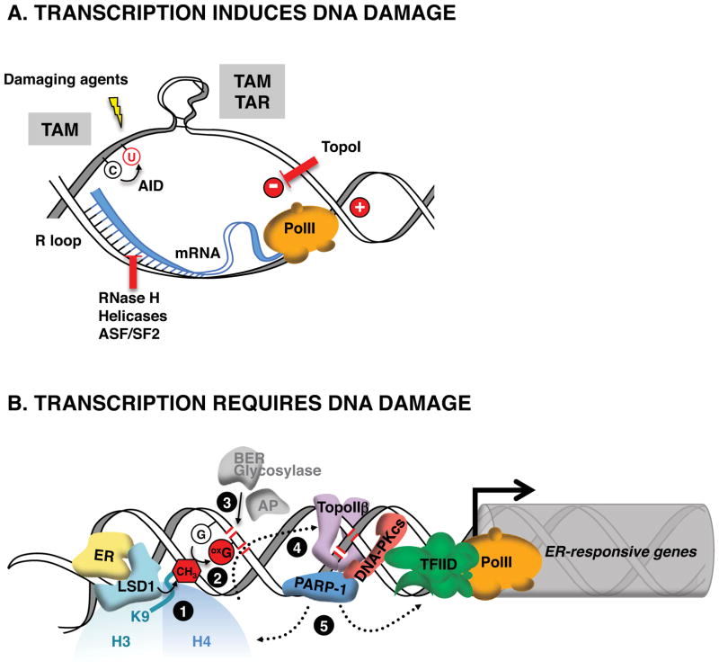
. The demethylation reaction releases reactive oxygen species that converts nearby guanines (G) into 8-oxo-guanines (oxG) formula image. oxG removal by base excision repair (BER) creates DNA nicks formula image that facilitate entrance of the endonuclease topoisomerase IIβ (TopoIIβ) formula image. TopoIIβ-induced double strand breaks recruit PARP-1 and DNA-PKcs repair enzymes, which induce a permissive chromatin architecture for transcription initiation formula image.

References

    1. Aguilera A. The connection between transcription and genomic instability. The EMBO journal. 2002;21:195–201. - PMC - PubMed
    1. Aguilera A, Garcia-Muse T. R loops: from transcription byproducts to threats to genome stability. Molecular cell. 2012;46:115–124. - PubMed
    1. Apostolou E, Ferrari F, Walsh RM, Bar-Nur O, Stadtfeld M, Cheloufi S, Stuart HT, Polo JM, Ohsumi TK, Borowsky ML, et al. Genome-wide chromatin interactions of the Nanog locus in pluripotency, differentiation, and reprogramming. Cell stem cell. 2013;12:699–712. - PMC - PubMed
    1. Araki M, Masutani C, Takemura M, Uchida A, Sugasawa K, Kondoh J, Ohkuma Y, Hanaoka F. Centrosome protein centrin 2/caltractin 1 is part of the xeroderma pigmentosum group C complex that initiates global genome nucleotide excision repair. The Journal of biological chemistry. 2001;276:18665–18672. - PubMed
    1. Barreto G, Schafer A, Marhold J, Stach D, Swaminathan SK, Handa V, Doderlein G, Maltry N, Wu W, Lyko F, Niehrs C. Gadd45a promotes epigenetic gene activation by repair-mediated DNA demethylation. Nature. 2007;445:671–675. - PubMed

Publication types

LinkOut - more resources