. 2013 Nov 7;52(3):434-46.
doi: 10.1016/j.molcel.2013.09.021. Epub 2013 Oct 24.

The DNA translocase FANCM/MHF promotes replication traverse of DNA interstrand crosslinks

Affiliations

The DNA translocase FANCM/MHF promotes replication traverse of DNA interstrand crosslinks

Jing Huang et al. Mol Cell. .

Abstract

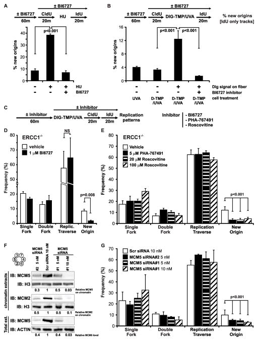
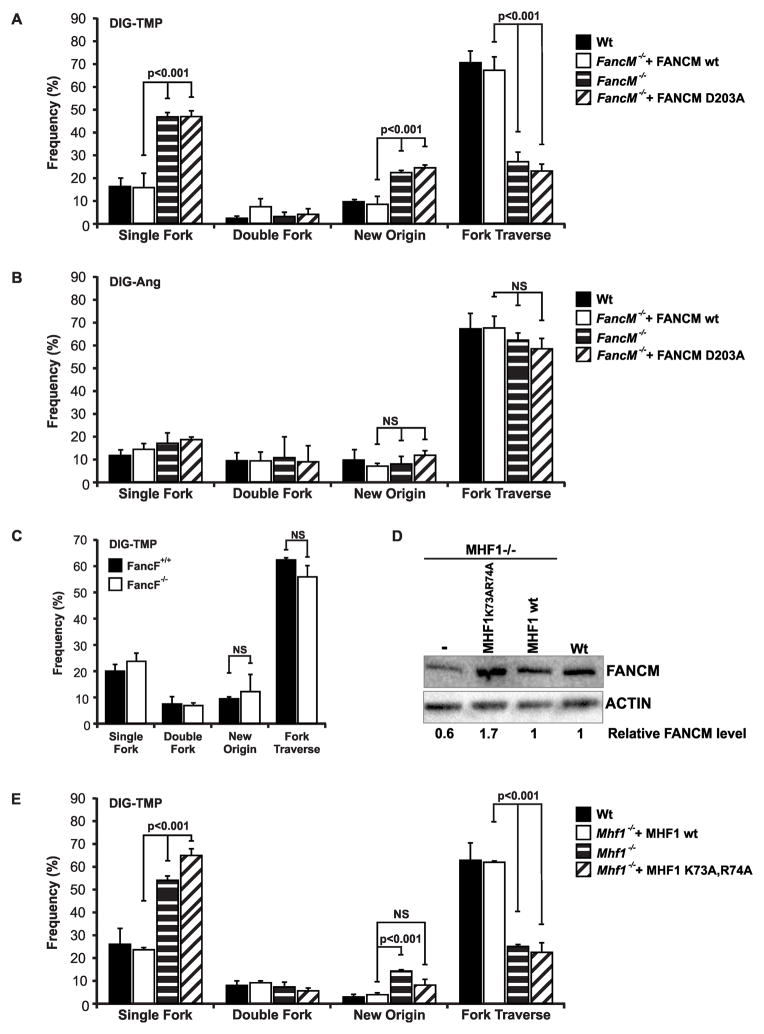
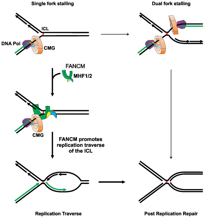
The replicative machinery encounters many impediments, some of which can be overcome by lesion bypass or replication restart pathways, leaving repair for a later time. However, interstrand crosslinks (ICLs), which preclude DNA unwinding, are considered absolute blocks to replication. Current models suggest that fork collisions, either from one or both sides of an ICL, initiate repair processes required for resumption of replication. To test these proposals, we developed a single-molecule technique for visualizing encounters of replication forks with ICLs as they occur in living cells. Surprisingly, the most frequent patterns were consistent with replication traverse of an ICL, without lesion repair. The traverse frequency was strongly reduced by inactivation of the translocase and DNA binding activities of the FANCM/MHF complex. The results indicate that translocase-based mechanisms enable DNA synthesis to continue past ICLs and that these lesions are not always absolute blocks to replication.


Figures

Fig. 1
Fig. 1. Cell treatment with Dig-TMP/UVA generates primarily ICLs
A. Dig-TMP, Dig-Ang. B. Sample preparation prior to analysis by LC-MS/MS. C. Determination of the ICL: MA ratio by LC-MS/MS analysis of genomic DNA (top) from cells treated with Dig-TMP/UVA and in a duplex oligodeoxyribonucleotide calibration standard (lower). Top left: the product-ion spectrum of the ESI-produced [M-3H]3− ion (m/z 757.1) of the tetranucleotide carrying the Dig-TMP ICL. Inset: the selected-ion chromatogram (SIC) for monitoring the m/z 757.1 →1096.4. Top right: the product-ion spectrum of the [M-2H]2− ion (m/z 818.5) of the dinucleotide carrying the Dig-TMP MA. Inset: the SIC for monitoring the m/z 818.5→1558.2 transition. The LC-MS/MS data for the standards are in the lower panels. Note the relative peak areas of the ICL and MAs in the SICs for the genomic DNA sample. See also Figures S2–S3.
Fig. 2
Fig. 2. Replication patterns in the vicinity of Dig-TMP and Dig-Ang adducts
A. Immunoquantum dot visualization of Dig-TMP (red) covalently linked to a DNA fiber labeled with CldU (green). B. Experimental design. C. Single and double sided replication patterns. D. Frequency of replication patterns in repair proficient cells. E. Frequency of single and double sided patterns as a function of pulse labeling time in wild type cells. F. Possible explanations for the double sided patterns. Dual fork collision, or partial repair of the ICL, could generate a single strand adduct which could be bypassed by the fork. For simplicity only the leading strand is depicted in green, but both the leading and lagging strands are labeled in the experiment. G. CHO cells were synchronized in G1 or S phase, treated with TMP/UVA and analyzed by an alkaline comet assay to determine the efficiency of unhooking (crosslink release). H. Pattern frequencies in ERCC1−/ − cells. I. Frequency of replication patterns as a function of pulse time in ERCC1−/ − cells. J. ICL: MA ratio in DNA harvested immediately after TMP/UVA treatment, and in replication tracts labeled with EdU for 1 hr. Data are presented as mean ±SD. See also Figure S4A, Table S1.
Fig. 3
Fig. 3. ICL status at the time of fork encounter
A. Experimental scheme. ERCC1−/ −cells were incubated with CldU (red) for several generations. Then the cells were incubated with IdU (green) for one cycle. The medium was changed and the cells treated with Dig-TMP/UVA, followed by incubation for 1 hr with EdU (blue). Cells were lysed and the DNA sheared prior to spreading on slides and immunostaining. The anticipated patterns are shown. B. Examples of daughter (blue) tracts associated with parental strands. The CldU tracts have been pseudo-colored purple to facilitate visualization of the Dig signal in the merged image. Blue tracts: without Dig-TMP (*); with a Dig-TMP MA signal on a green fiber (**); with a Dig-TMP ICL on a purple/green fiber (***). C. Distribution of EdU (blue) tracts on fibers from ERCC1−/ − cells, or repair proficient wild type cells, treated with Dig-TMP/UVA, Dig-Ang/UVA, or UVA only. The two bars on the right represent fibers without, or with, Dig-TMP signals from cells treated with Dig-TMP/UVA. Data are presented as mean ±SD. See also Figure S4B, Table S2.
Fig. 4
Fig. 4. Replication traverse of ICLs
A. Sequential labeling defines the direction of replication in the vicinity of ICLs. Cells were treated with Dig-TMP/UVA and then pulsed for 20 min with CldU (purple), followed by a 20 min pulse of IdU (green). Pseudo-coloring as in Fig 3. B, C, D, E. Images and interpretation of replication patterns in the vicinity of Dig-TMP signals. Another pattern (not shown), in which a Dig-TMP signal appeared in a new origin (purple flanked by green), appeared at a frequency of less than 1 %. F. Quantitation of replication patterns in Wt and ERCC1−/ − cells, mean ±SD. G. Time cost of replication traverse. The lengths of IdU tracts, with or without a Dig-TMP signal, from cells treated with Dig-TMP, were measured and the time difference calculated. The same analysis was performed with IdU tracts from cells treated with Dig-Ang. See also Figure S5, Table S3.
Fig. 5
Fig. 5. Dormant origin activation does not explain the traverse patterns
A. The PLK-1 inhibitor BI6727 blocks dormant origin activation induced by HU treatment. After a 20 min CldU pulse, cells were incubated for 12 hours in the presence of 2 mM HU, followed by an IdU pulse. Experiments were done in parallel with or without BI6727 (Methods). B. Dormant origin activation by Dig-TMP/UVA treatment is blocked by BI6727. C, D, E. The frequency of fork traverse patterns is not affected by the inhibitors BI6727, PHA-767491, or roscovitine. Cells were treated with Dig-TMP/UVA, followed by sequential pulses of CldU and IdU, all in the presence of drugs or vehicle. F. Western blot analysis of MCM5 and MCM2 in total cell extracts or in chromatin, following treatment of cells with siRNAs against MCM5. G. The frequency of fork traverse patterns is unaffected by knockdown of MCM5. Data are presented as mean ±SD (p<0.001, chi-square test). See also Figure S6, Table S4.
Fig. 6
Fig. 6. Replication traverse of ICLs, but not MAs, is promoted by FANCM translocase activity
A. Replication fork traverse of ICLs is reduced in FANCM−/ − cells. DT40 wild type or FANCM−/ −, or FANCM−/ − cells expressing wild type FANCM, or a translocase mutant version (D203A), were treated with Dig-TMP/UVA followed by sequential pulses of CldU and IdU. B. FANCM deficiency does not influence the frequency of replication patterns in cells treated with Dig-Ang. C. FANCF deficiency does not influence the frequency of replication patterns in cells treated with Dig-TMP. (D) Relative FANCM protein levels in MHF1−/ − cells and complemented cells total cell extracts. E. Traverse patterns are reduced in cells deficient in MHF1 or expressing a DNA binding mutant of MHF1(K73A,R74A). Data are presented as mean ±SD (p<0.001, chi-square test). See also Figure S7, Table S5.
Fig. 7
Fig. 7. Replication traverse of ICLs
Following the encounter of a single replication fork, a second fork may collide with the same ICL from the other side (Raschle et al., 2008). More frequently, recruitment of FANCM/MHF1/2 to the stalled fork is followed by replication restart on the distal side of the ICL. After Okazaki fragment ligation the DNA structures formed by either pathway are identical.

References

    1. Bagby GC, Alter BP. Fanconi anemia. Semin Hematol. 2006;43:147–156. - PubMed
    1. Bakker ST, van de Vrugt HJ, Rooimans MA, Oostra AB, Steltenpool J, Delzenne-Goette E, van der Wal A, van dV, Joenje H, te RH, de Winter JP. Fancm-deficient mice reveal unique features of Fanconi anemia complementation group M. Hum Mol Genet. 2009;18:3484–3495. - PubMed
    1. Bastia D, Zzaman S, Krings G, Saxena M, Peng X, Greenberg MM. Replication termination mechanism as revealed by Tus-mediated polar arrest of a sliding helicase. Proc Natl Acad Sci U S A. 2008;105:12831–12836. - PMC - PubMed
    1. Blackford AN, Schwab RA, Nieminuszczy J, Deans AJ, West SC, Niedzwiedz W. The DNA translocase activity of FANCM protects stalled replication forks. Hum Mol Genet. 2012;21:2005–2016. - PubMed
    1. Blow JJ, Ge XQ. A model for DNA replication showing how dormant origins safeguard against replication fork failure. EMBO Rep. 2009;10:406–412. - PMC - PubMed

Publication types