. 2013 Nov 21;52(4):541-53.
doi: 10.1016/j.molcel.2013.09.025. Epub 2013 Oct 24.

PrimPol, an archaic primase/polymerase operating in human cells

Affiliations

PrimPol, an archaic primase/polymerase operating in human cells

Sara García-Gómez et al. Mol Cell. .

Abstract

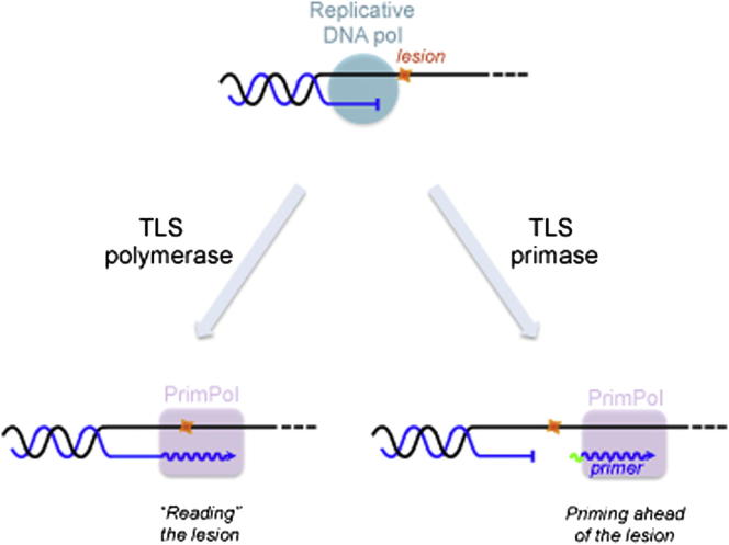
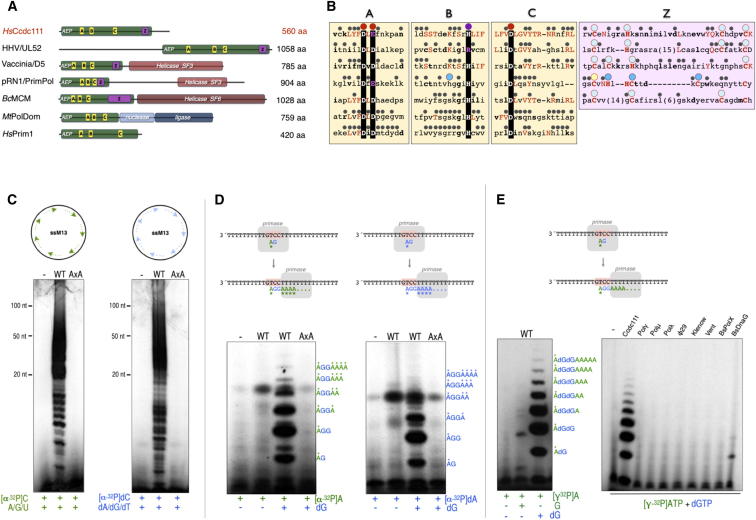
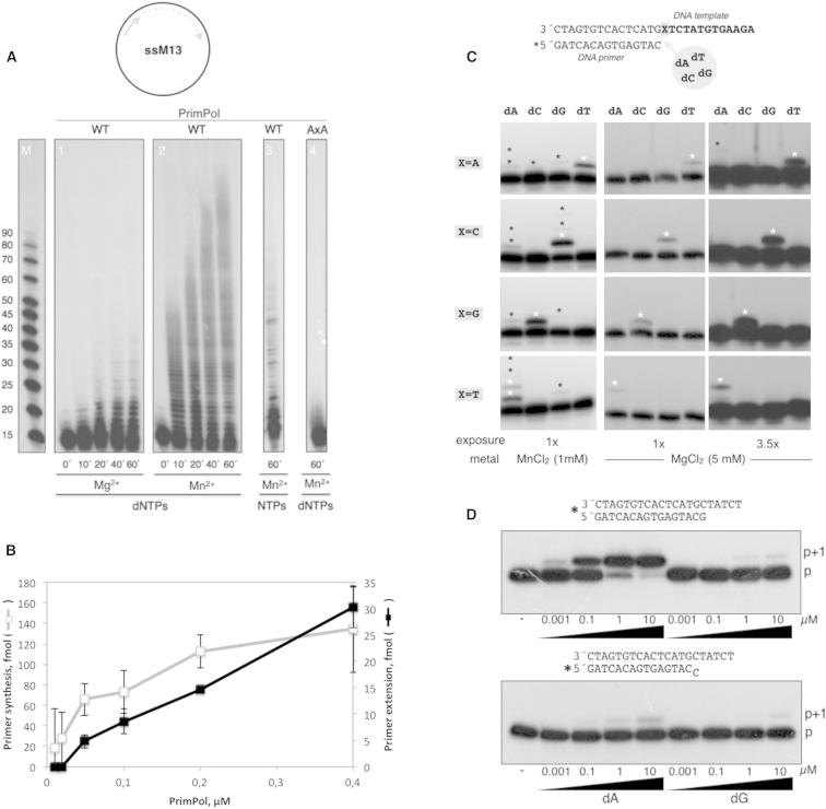
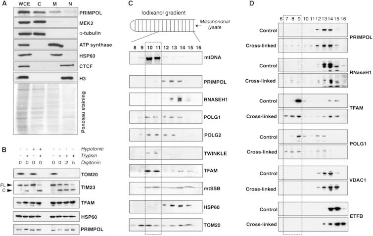
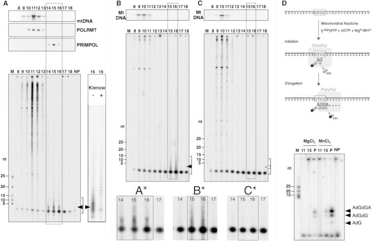
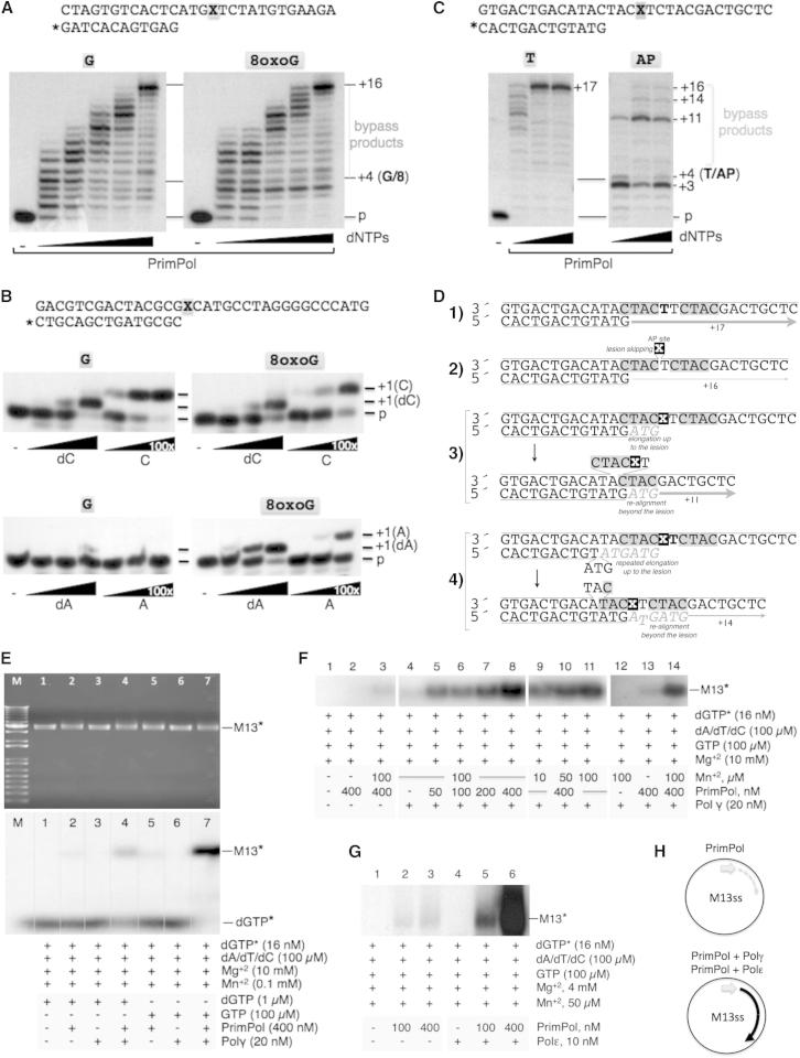
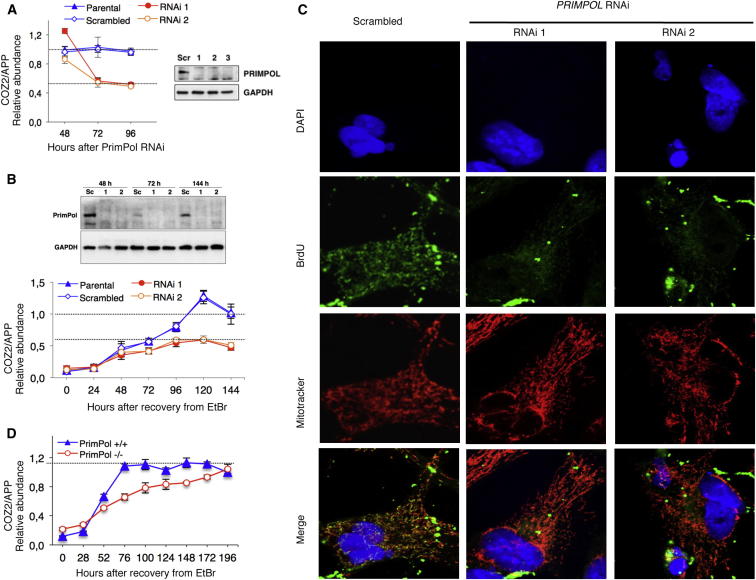
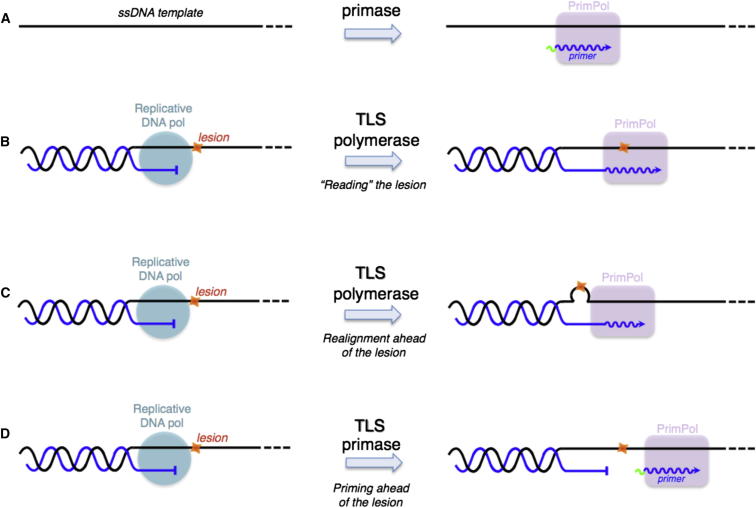
We describe a second primase in human cells, PrimPol, which has the ability to start DNA chains with deoxynucleotides unlike regular primases, which use exclusively ribonucleotides. Moreover, PrimPol is also a DNA polymerase tailored to bypass the most common oxidative lesions in DNA, such as abasic sites and 8-oxoguanine. Subcellular fractionation and immunodetection studies indicated that PrimPol is present in both nuclear and mitochondrial DNA compartments. PrimPol activity is detectable in mitochondrial lysates from human and mouse cells but is absent from mitochondria derived from PRIMPOL knockout mice. PRIMPOL gene silencing or ablation in human and mouse cells impaired mitochondrial DNA replication. On the basis of the synergy observed with replicative DNA polymerases Polγ and Polε, PrimPol is proposed to facilitate replication fork progression by acting as a translesion DNA polymerase or as a specific DNA primase reinitiating downstream of lesions that block synthesis during both mitochondrial and nuclear DNA replication.


Figures

None
Graphical abstract
Figure 1
Figure 1
Human ccdc111 Is a Unique RNA/DNA Primase (A) Modular organization of various AEP-like enzymes. A conserved AEP domain (green bar) contains the three conserved regions A, B, and C, which form the primase active site. Hsccdc111 contains, in addition to regions A, B, and C, a Zn-finger-containing region also present in the viral, plasmidic, and bacterial specimens. Additionally, some APE enzymes contain additional activities and domains as helicases (red bars) or a combination of nuclease and ligase (blue bars). Hsccdc111, human ccdc111 protein; HHV/UL52, herpes virus UL52 primase; Vaccinia/D5, vaccinia virus primase; pRN1/PrimPol, plasmid pRN1 ORF904 from S. islandicus; BcMCM, PrimPol helicase from Bacillus cereus; MtPolDom, LigD polymerization domain from Mycobacterium tuberculosis; and HsPrim1, small catalytic subunit of the human RNA primase. See also Figure S1. (B) Conserved regions in proteins having and AEP-related domain. Large dots indicate metal ligands (red), nucleotide ligands (purple), and demonstrated (cyan) or potential (light blue) cysteine residues forming a Zn finger. Small dots indicate invariant residues in each group of AEP-related enzymes. Residues that are either conserved or identical to those in ccdc111 are indicated in bold or red letters, respectively. See also Figure S1 for a more extensive alignment. (C) Primer synthesis on M13 ssDNA using either NTPs or dNTPs. A scheme of the primase reaction is shown. WT ccdc111 (but not mutant AxA) synthesized de novo RNA (left) or DNA (right) primers on the M13 ssDNA circular template in the presence of activating Mn2+ ions. See also Figures S2 and S3. (D) Priming at an ori-containing oligonucleotide. The schematic shows the different (initiation and elongation) products synthesized by a primase that recognizes the central GTCC sequence as a starting ori, providing the indicated combinations of NTPs (in green) and dNTPs (in blue). The primase products obtained with ccdc111 were very similar when either ATP (left) or dATP (right) were provided as 5′ nucleotide (in addition to dGTP, provided as a common 3′nucleotide). See also Figures S2 and S3. (E) The same primase assay as in (D) except that primase products were labeled with [γ-32P]ATP as the 5′ nucleotide. dGTP was a much better alternative than GTP as 3′ nucleotide (left). In these preferred conditions, the formation of the ribo-deoxy dimer (and further elongation products) is a hallmark of PrimPol, which cannot be obtained with other DNA polymerases or conventional primases (right).
Figure 2
Figure 2
ccdc111 Is a DNA-Dependent DNA Polymerase (A) Elongation of a 17-mer primer annealed to M13 circular ssDNA in the presence of either 5 mM MgCl2 or 1 mM MnCl2 as metal activators. WT ccdc111 (but not mutant AxA) extended the primer producing elongation intermediates characteristic of distributive DNA synthesis. NTPs were worse substrates for polymerization by ccdc111. (B) Compared efficiency of primase and polymerase activities of PrimPol. The primase assay, using the 29-mer GTCC oligo (5′-T15CCTGT10-3′) as template, and the DNA polymerase assay, using a template/primer oligonucleotide, were carried out as described in the Experimental Procedures. Data are represented as mean ± SD (n = 3). See also Figure S3. (C) Matched versus mismatched nucleotide insertion at the four template bases. The four template/primer structures used, differing in the first template base (X), are indicated. Nucleotide insertion on each template/primer was analyzed in the presence of each individual dNTP. PrimPol preferentially inserted the complementary nucleotide dictated by the first available templating base. The insertion of some errors (indicated with asterisks) can be detected. (D) Polymerization assays were carried out with either a matched (top) or mismatched (middle) template/primer and the indicated concentrations of a correct (dA) or wrong (dG) nucleotide. After incubation, +1 extension of the 5′-labeled () strand was analyzed by denaturing gel and autoradiography.
Figure 3
Figure 3
PrimPol Localizes at Both DNA Compartments, Mitochondria, and Nucleus (A) 5 × 106 HeLa cells were fractionated (see the Experimental Procedures) in order to estimate the distribution of PrimPol between the different subcellular compartments. The distribution, calculated as an average of three independent experiments, was 47% cytosolic (C), 34% mitochondrial (M), and 19% nuclear (N). WCE, whole-cell extract. Mek2 and tubulin are shown as controls for the cytosolic fraction, Hsp60 and ATP synthase are shown as controls for the mitochondrial fraction, and CTCF and H3 are shown as controls for the nuclear fraction. (B) Mitochondria isolated from HEK 293T cells were treated with hypotonic buffer or increasing concentrations of digitonin (2 or 5 ×10 mg/ml) followed or not by tripsin digestion. The amount of PrimPol remaining after each treatment was determined by immunoblot. TOM20 and TIM23 were used as outer and inner membrane markers, whereas TFAM and HSP60 were used as matrix markers. (C) Mitochondria isolated from HEK 293T cells were subjected to lysis and fractionation on an iodixanol gradient (IG). DNA and protein were recovered from the different fractions and hybridized to a DNA probe specific for human mtDNA or antibodies against PrimPol and proteins involved in mtDNA maintenance, respectively. (D) Mitochondria isolated from HEK 293T cells were crosslinked with formaldehyde or left untreated and subjected to lysis and fractionation on an IG. Proteins were recovered from the different fractions and hybridized to antibodies against PrimPol, proteins involved in mtDNA maintenance (RHaseH1, TFAM, and POLG1), and controls for nonspecific crosslinking (VDAC1 and ETFB).
Figure 4
Figure 4
PrimPol-Enriched Fractions from Mitochondria Have Primase Activity Similar to the Purified Protein (A) Mitochondria isolated from HEK 293T cells were treated with trypsin prior to lysis and fractionation on an IG. Primase assays were carried out from each sample with oligo dT25 as template, and the resulting products were resolved in 7M urea-containing 8% polyacrylamide gels. Filled arrowheads indicate specific products detected in PrimPol fractions. The presence of mtDNA was confirmed by southern blot, whereas PrimPol and POLRMT were detected by immunoblotting. (B and C) Mitochondria isolated from PrimPol +/+ (B) or PrimPol −/− (C) MEFs (see also Figure S4) were fractionated in IG, and primase assays were carried out as in (A). Filled or empty arrowheads indicate specific products detected in PrimPol fractions or their absence, respectively. Zoom-in regions (A, B, and C) contain PrimPol specific products from (A), (B), and (C), respectively. (D) PrimPol-specific labeling of de novo initiation products with oligo d(T20GTCCT36) as a template. Selected mtDNA-, POLRMT- (11), and PrimPol-enriched (15) IG fractions were assayed in parallel to recombinant purified PrimPol (P) and no-protein control (NP) in the conditions described in Figure S2E. The resulting products were resolved in 8M urea-containing 20% polyacrylamide gels and visualized by autoradiography.
Figure 5
Figure 5
PrimPol Tolerates 8oxoG and Abasic Sites in DNA and Functionally Cooperates with Polγ and Polε (A) Running start DNA polymerization by human PrimPol on a control template (left) or on a template containing an 8oxoG lesion (right). dNTPs were provided at 0.05, 0.1, 0.5, 1, or 10 μM. The position of the lesion in the template strand is marked (X) in the top schematic as well as in the autoradiogram. Human PrimPol elongated the damaged template with the same efficiency as in the case of the undamaged template. See also Figure S5 for a comparison to Polγ. (B) Standing start DNA polymerization by human PrimPol on a control template (left) or on a template containing an 8oxoG lesion (right). The error-free insertion of either dCTP or CTP and the error-prone insertion of either dATP or ATP are compared at the indicated concentrations. The mobility of the +1 extended (opposite the lesion) primers is indicated. (C) Running start DNA polymerization assay on a template containing an AP site. dNTPs were provided at 1, 10, and 100 μM. Human PrimPol bypassed a significant proportion of the templates containing the AP site (right) but generated incomplete bypass products. See also Figure S5 for a comparison with Polγ. (D) Schematics summarizing the origin and relative abundance of different bypass products shown in (C): (1) efficient copy of the normal template (+17), (2) polymerization + skipping of the AP nucleotide (+16), (3) elongation up to the lesion + realignment beyond the lesion (+11), and (4) elongation and primer slippage before the lesion + realignment beyond the lesion (+14). (E) Synergy between human PrimPol and human Polγ. Using circular M13 ssDNA as template and the indicated amounts of both enzymes (see the Experimental Procedures), the RNA/DNA primers made by PrimPol were readily extended by Polγ. The PrimPol-Polγ synergistic effect was maximal when GTP was provided in order to maximize the primase activity of PrimPol. (F) Synergy with Polγ allowed the detection of PrimPol action at much lower enzyme (50 nM) and manganese (10 μm) concentrations. (G) Synergy between human PrimPol and human Polε. The DNA primers made by PrimPol on M13 ssDNA were efficiently extended by Polγ. (D) Schematics of the functional coupling between PrimPol and the replicative DNA polymerases Polγ and Polε.
Figure 6
Figure 6
PRIMPOL Gene Silencing or Ablation Perturbs mtDNA Replication (A) HEK 293T cells were transfected with PRIMPOL-specific dsRNAs (see the Experimental Procedures), and DNA was harvested daily from 48 to 96 hr after transfection. Controls were untransfected parental and scrambled dsRNA transfected HEK 293T cells. mtDNA copy number was assayed by quantitative PCR (qPCR; see the Experimental Procedures). Data are represented as mean ± SEM. Proteins from cells harvested 72 hr after transfection were immunoblotted for specific antibodies against PrimPol and GAPDH as controls (right). See also Figure S6. (B) HEK 293T cells were depleted of mtDNA to 10% of normal by exposure to 100 ng/ml EtBr for 72 hr. After 24 hr of depletion, they were transfected with scrambled or two PRIMPOL-specific dsRNAs (RNAi 1 and RNAi 2), and DNA was harvested daily from 0 to 192 hr after removal of the EtBr. mtDNA copy number was assayed by qPCR (see the Experimental Procedures). Data are represented as mean ± SEM. Protein was also harvested at 48, 72, and 144 hr after the removal of the EtBr. PrimPol and GAPDH (reference protein) were detected by immunoblotting with specific antibodies. (C) HOS cells, transfected with scrambled or two PRIMPOL-specific dsRNAs (RNAi 1 and RNAi 2) for 72 hr, were incubated with BrdU for 24 hr in the presence of aphidicolin. After fixation, cells were incubated with DAPI (blue), an antibody against BrdU (green), and MitoTracker (red) and examined by confocal microscopy. (D) PRIMPOL+/+ and PRIMPOL−/− MEFs (see also Figure S4) were depleted of mtDNA to 10%–20% of normal by exposure to 200 ng/ml EtBr for 172 hr. DNA was harvested daily from 0 to 196 hr after the removal of the EtBr. mtDNA copy number was assayed by qPCR (see the Experimental Procedures). Data are represented as mean ± SEM.
Figure 7
Figure 7
Alternative Functions of PrimPol in DNA Replication (A) PrimPol can act as a “conventional” primase but is able to start DNA synthesis on its own. (B) PrimPol can act as a “classical” TLS polymerase, reading lesions as 8oxoG. (C) PrimPol can realign the stalled primer terminus ahead of the lesion, acting as a “pseudo” TLS polymerase. (D) PrimPol can reinitiate DNA synthesis ahead of the lesion, by means of its DNA primase activity, at the single-stranded region generated by continued helix opening after the replicative DNA polymerase got stalled at a lesion. Such a “TLS primase” activity would allow replication fork progression, but a lesion-containing gap would be left behind for later repair.

References

    1. Aravind L., Leipe D.D., Koonin E.V. Toprim—a conserved catalytic domain in type IA and II topoisomerases, DnaG-type primases, OLD family nucleases and RecR proteins. Nucleic Acids Res. 1998;26:4205–4213. - PMC - PubMed
    1. Bernad A., Blanco L., Lázaro J.M., Martín G., Salas M. A conserved 3′----5′ exonuclease active site in prokaryotic and eukaryotic DNA polymerases. Cell. 1989;59:219–228. - PubMed
    1. Berquist B.R., Wilson D.M., 3rd Pathways for repairing and tolerating the spectrum of oxidative DNA lesions. Cancer Lett. 2012;327:61–72. - PMC - PubMed
    1. Blanca G., Shevelev I., Ramadan K., Villani G., Spadari S., Hübscher U., Maga G. Human DNA polymerase lambda diverged in evolution from DNA polymerase beta toward specific Mn(++) dependence: a kinetic and thermodynamic study. Biochemistry. 2003;42:7467–7476. - PubMed
    1. Bolden A., Noy G.P., Weissbach A. DNA polymerase of mitochondria is a gamma-polymerase. J. Biol. Chem. 1977;252:3351–3356. - PubMed

Publication types

MeSH terms