. 2013 Dec 3;21(12):2119-30.
doi: 10.1016/j.str.2013.09.008. Epub 2013 Oct 24.

Structural dynamics and topology of phosphorylated phospholamban homopentamer reveal its role in the regulation of calcium transport

Affiliations

Structural dynamics and topology of phosphorylated phospholamban homopentamer reveal its role in the regulation of calcium transport

Vitaly V Vostrikov et al. Structure. .

Abstract

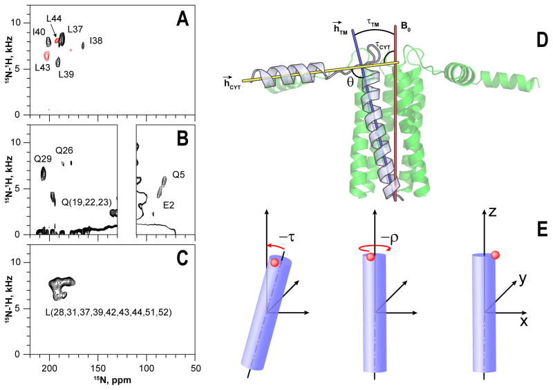
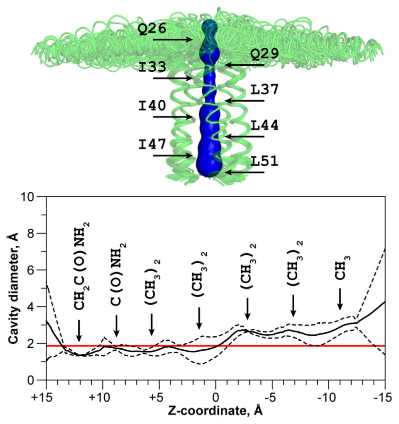
Phospholamban (PLN) inhibits the sarco(endo)plasmic reticulum Ca²⁺-ATPase (SERCA), thereby regulating cardiac diastole. In membranes, PLN assembles into homopentamers that in both the phosphorylated and nonphosphorylated states have been proposed to form ion-selective channels. Here, we determined the structure of the phosphorylated pentamer using a combination of solution and solid-state nuclear magnetic resonance methods. We found that the pinwheel architecture of the homopentamer is preserved upon phosphorylation, with each monomer having an L-shaped conformation. The TM domains form a hydrophobic pore approximately 24 Å long and 2 Å in diameter, which is inconsistent with canonical Ca²⁺-selective channels. Phosphorylation, however, enhances the conformational dynamics of the cytoplasmic region of PLN, causing partial unwinding of the amphipathic helix. We propose that PLN oligomers act as storage for active monomers, keeping SERCA function within a physiological window.


Figures

Figure 1
Figure 1
PISEMA spectra of the selectively labeled pS16-PLNWT in mechanically aligned DOPC:DOPE lipid bilayers. Samples were prepared with synthetic (A) or recombinant (B–C) proteins. Two spectra obtained from different synthetic samples are color coded in panel A. Definition of topological angles τ (tilt), ρ (rotation), θ (angle between helices) (D) and their calculation from the structural data (E). Structural model of pS16-PLNWT pentamer, illustrating the tilt angles of the transmembrane (τTM) and cytoplasmic (τCYT) helices defined as the angles between the applied magnetic field (B0) and the corresponding α-helix vectors (h→). The inter-domain angle θ is defined between the vectors h→TM and h→CYT. Performing successive right-handed rotations of an α-helix by negative τ and negative ρ aligns the reference atom (blue) with the X-axis (see Table 1). Helical axes were calculated using the average direction of carbonyl bonds, employing script from PyMOL repository.
Figure 2
Figure 2
Arrangement of Leu (red) and Ile (green) side chains on the structure of pS16 form (one protomer is removed for clarity) (A). Strips from DARR spectrum of the mixed Ile and Leu pS16-PLNWT (each protomer labeled with only one amino acid type) (B). Ambiguous restraints modeled from the DARR data (C).
Figure 3
Figure 3
Structural ensembles of PLNWT in the non-phosphorylated (left) and pS16 (right) forms as viewed along the membrane normal (A, B) or perpendicular to it (E, F). Structures are aligned on all backbone atoms. The residues 1–17 are shown in ribbon projection, illustrating disorder near the phosphorylation site when the alignment is performed on the heavy atoms of only these residues (C, D). Side chains of the cytoplasmic domain are shown as sticks. Carbon atoms of the side chains are colored green for hydrophobic residues and cyan for hydrophilic ones. Structural statistics are reported in the Supplemental Information (Tables S1–S3, Figures S7–S8).
Figure 4
Figure 4
Coupled enzyme assays for SERCA alone (black), SERCA in the presence of PLNWT (red) or pS16-PLNWT (blue).
Figure 5
Figure 5
Model independent relaxation parameters for non-phosphorylated and pS16 forms of pentameric PLNWT and monomeric PLNAFA at 600 MHz (A, C). Model independent relaxation parameters derived from the 600 MHz data plotted as a color map on the structures of PLN in the non-phosphorylated and pS16 forms (B, D). Color scales for a given parameter are indicated and ramped in increment of one unit, absent data is in white. See also Figure S6.
Figure 6
Figure 6
Inner pore of the pentameric assembly of pS16-PLNWT. The structural features were analyzed with the program MOLE (Petrek et al., 2007). The surface of the pore is shown in blue and is overlaid with the structural ensemble, aligned at the transmembrane domains. The maximum pore diameter has been calculated for the five lowest energy structures and is presented in the mean ± standard deviation form. Red line indicates the diameter of non-hydrated Ca2+ ion (1.98 Å, (Tsien et al., 1987)).

References

    1. Abu-Baker S, Lorigan GA. Phospholamban and its phosphorylated form interact differently with lipid bilayers: a 31P, 2H, and 13C solid-state NMR spectroscopic study. Biochemistry. 2006;45:13312–13322. - PMC - PubMed
    1. Anderson ME, Brown JH, Bers DM. CaMKII in myocardial hypertrophy and heart failure. J Mol Cell Cardiol. 2011;51:468–473. - PMC - PubMed
    1. Aschar-Sobbi R, Emmett TL, Kargacin GJ, Kargacin ME. Phospholamban phosphorylation increases the passive calcium leak from cardiac sarcoplasmic reticulum. Pflugers Arch. 2012;464:295–305. - PubMed
    1. Becucci L, Cembran A, Karim CB, Thomas DD, Guidelli R, Gao J, Veglia G. On the function of pentameric phospholamban: ion channel or storage form? Biophys J. 2009;96:L60–L62. - PMC - PubMed
    1. Becucci L, Foresti ML, Schwan A, Guidelli R. Can proton pumping by SERCA enhance the regulatory role of phospholamban and sarcolipin? Biochim Biophys Acta. 2013;1828:2682–2690. - PubMed

Publication types

MeSH terms

Substances

Associated data

LinkOut - more resources