. 2013 Nov 7;155(4):909-921.
doi: 10.1016/j.cell.2013.10.023.

A zebrafish embryo culture system defines factors that promote vertebrate myogenesis across species

Affiliations

A zebrafish embryo culture system defines factors that promote vertebrate myogenesis across species

Cong Xu et al. Cell. .

Abstract

Ex vivo expansion of satellite cells and directed differentiation of pluripotent cells to mature skeletal muscle have proved difficult challenges for regenerative biology. Using a zebrafish embryo culture system with reporters of early and late skeletal muscle differentiation, we examined the influence of 2,400 chemicals on myogenesis and identified six that expanded muscle progenitors, including three GSK3β inhibitors, two calpain inhibitors, and one adenylyl cyclase activator, forskolin. Forskolin also enhanced proliferation of mouse satellite cells in culture and maintained their ability to engraft muscle in vivo. A combination of bFGF, forskolin, and the GSK3β inhibitor BIO induced skeletal muscle differentiation in human induced pluripotent stem cells (iPSCs) and produced engraftable myogenic progenitors that contributed to muscle repair in vivo. In summary, these studies reveal functionally conserved pathways regulating myogenesis across species and identify chemical compounds that expand mouse satellite cells and differentiate human iPSCs into engraftable muscle.


Figures

Figure 1
Figure 1. A chemical genetic screen to identify modifiers of skeletal muscle development
(A) myf5-GFP;mylz2-mCherry double transgenic expression recapitulates expression of the endogenous genes. myf5-GFPis first detected at the 11-somite stage. mylz2-mCherry expression is not observed until 32 hpf. Scale bars represent 200 μm. (B) myf5-GFP;mylz2-mCherry embryos were dissociated at the oblong stage and cultured in zESC medium. Images were taken 48 hours after plating. Scale bars represent 250 μm. (C)Expression of myogenic genes measured by qRT-PCR. Blastomere cells were cultured and harvested at 24 hours. Error bars represent standard deviation (SD) from the triplicate reactions. All values are normalized to β-actin and expression data of basic medium (grey bars, lacking bFGF)are set to 1. (D) Expression of myogenic genes by different populations of blastomere cells isolated by FACS after 24 hour culture with bFGF. Error bars represent standard deviation (SD) from triplicate reactions. Expression data are normalized to β-actin and expression values of double negative population (blue bars) are set to 1. See also Figure S1.
Figure 2
Figure 2. Chemical genetic screens to identify modifiers of skeletal muscle development
(A) Schematic of a high-throughput image-based chemical screening assay. Approximately 800 myf5-GFP;mylz2-mCherry double transgenic embryos were collected and dissociated at the oblong stage. Resulting blastomere cells were aliquotted into 4384-well plates with pre-added chemicals. After 2 days, the 384-well plates were imaged and analzyed using a Celigo cytometer. (B) Sample images from the modifier screen. Hits could be grouped into three categories. Category I has only myf5-GFP expression. Category II has a decreased amount of both fluorescent colors. Category III has increased expression of both. Scale bars represent 250 μm. (C) Hits from the enhancer screen. 6 chemicals that increase the GFP and mCherry signals were identified. Scale bars represent 250 μM. See also Figure S2 and Tables S1 and S2.
Figure 3
Figure 3. Forskolin treatment elevates cAMP level and increases proliferation of satellite cells from both healthy and dystrophic mice
(A) Satellite cells from C57BL/6J mice were cultured and treated with DMSO or forskolin in the absence or presence of bFGF, as indicated. Total cell number was determined after 5 days (mean +/− SEM, n=4). Data presented as fold change, normalized to“DMSO no bFGF” controls. (B) Fold change in cell number from cultures of mdx satellite cells after 5 days in vitro with bFGF alone, DMSO + bFGF, or forskolin+ bFGF(mean +/− SEM, n=4). Data normalized to “DMSO +bFGF” controls. (C) Concentration of cAMP in cultured satellite cells is increased after treatment with 25 μM, 50 μM or 100 μM of forskolin, as compared to the DMSO treated cells (mean +/− SD, n=5). Data normalized “DMSO” controls. (D) Experimental scheme for myogenic colony forming assay. Satellite cells were isolated from C57BL/6J mice and a single cell was plated into each well of 96-well plates. Cells were cultured for 6 days and treated with DMSO or forskolin. Number of wells containing a myogenic colony and number of cells in each colony were counted after 6 days. (E) Clonal plating efficiency of satellite cells is not altered by forskolin treatment, as compared to DMSO-treated cells (mean +/− SD, n=4). (F) Forskolin treatment increases the number of myogenic cells arising from a single satellite cell in each well, showing an increase in cell proliferation. Data are plotted as the % of colonies containing the indicated number of cells (mean +/− SD, n=4).★: P <0.05, ★★: P<0.01 See also Figure S3.
Figure 4
Figure 4. Forskolin-treated satellite cells exhibit effective differentiation in vitro
(A) Experimental scheme. Satellite cells from C57BL/6J mice were cultured in the presence of bFGF for 5 days. Cells were harvested on day 5 and equal numbers of cells were induced to differentiate in the presence of forskolin or DMSO. (B) Images of satellite cells differentiated in the presence of DMSO (left) or forskolin (right) and stained for Myosin Heavy Chain (MHC, red) and nuclei (blue). Scale bars represent 200 μm. (C) Quantification of percentage of nuclei in myotubes after satellite cell differentiation in the presence of forskolin or DMSO (mean +/− SEM, n=4). Differentiation potential of satellite cells is unaffected by forskolin (p=non-significant (NS)). (D) Satellite cells from C57BL/6J mice were cultured with bFGF and forskolin/DMSO for 5 days. Cells were harvested on day 5 and equal numbers of cells were induced to differentiate after removal of the compound. (E) Images of DMSO (left) or forskolin (right) treated satellite cells differentiated after removal of compound and stained for MHC (red) and nuclei (blue). Scale bars represent 200 μm (F) Quantification of percentage of nuclei in myotubes after differentiation of forskolin or DMSO treated cells (mean +/− SEM, n=5). Forskolin-treated satellite cells show no defect in myotube formation. See also Figure S4.
Figure 5
Figure 5. Forskolin-treated cultured satellite cells retain immunophenotypic characteristics of freshly isolated satellite cells and engraft skeletal muscle in vivo
(A) Representative FACS plots depict CD45SCA-1MAC1 cells gated for the CXCR4+ and β1 Integrin+ subset of freshly isolated (left panel) and cultured satellite cells (initially sorted as 100% CXCR4+ and β-1 Integrin+) treated with DMSO (middle panel) or forskolin (right panel). (B) Average frequency (mean +/− SEM, n=6) of CXCR4+β1-Integrin+ cells among cultured satellite cells treated with DMSO or forskolin, quantified by FACS. Most cultured satellite cells treated with either DMSO or forskolin retain expression of CXCR4 and β1 Integrin. (C) Experimental scheme. GFP+ satellite cells were harvested from β-actin-GFP mice and transplanted into the TA muscle of recipient mdx mice, injured 1 day previously by injection of cardiotoxin. Cells were transplanted either immediately after isolation or following five days in culture with DMSO or forskolin treatment. (D) Total number of cultured GFP+ satellite cells that were obtained from 6000 freshly isolated cells and used for transplantation into mdx muscle. Cell number was, on average, 2.5 times greater in forskolin-treated as compared to DMSO treated cultures (mean +/− SD, n=6). (E) Transverse frozen section of TA muscle from mdx mice transplanted with 6000 freshly isolated satellite cells (left panel), cultured DMSO-treated satellite cells expanded from 6000 freshly isolated cells (middle panel) or cultured forskolin-treated satellite cells expanded from 6000 freshly isolated cells (right panel). Laminin staining is shown in red. Scale bars represent 200 μm. (F) Transverse frozen sections of TA muscle from mdx mice transplanted with 200,000 cultured DMSO-treated (left) or 200,000 cultured forskolin-treated (right) GFP+ satellite cells. Laminin staining is shown in red. Scale bars represent 200 μm. (G) Quantification of donor derived (GFP+) myofibers in mdx muscles transplanted with freshly isolated, DMSO-cultured or forskolin-cultured satellite cells (mean +/− SD, n=6). (H) Quantification of donor derived myofibers in mdx muscle transplanted with 200,000 cultured DMSO-treated or 200,000 cultured forskolin-treated satellite cells (mean +/− SD, n=4).
Figure 6
Figure 6. Treatment of human iPSCs with muscle promoting cocktail induces skeletal muscle differentiation
(A, B) Gene expression analysis of EBs (A) and monolayer cells (B). Cells were harvested at the times indicated for RNA extraction. Ectodermal, endodermal and mesodermal genes were analyzed by quantitative RT-PCR using GAPDH as a housekeeping gene. Changes in gene expression are indicated relative to undifferentiated iPSCs (Day 0). Bars represent the standard deviation of three independent experiments (*p<0.05, **<0.01, ***<0.001). (C)Immunostaining of differentiated iPSCs. Under terminal differentiating procedures (day 36) most of the cells express Desmin (red) and Myogenin (green), forming multinucleated myofibers. Cells were also stained with Hoechst (blue). The number on each panel represents the percentage of cells expressing Desmin and Myogenin. Scale bars represent 100μm. (D) Representative electron microscopy image of differentiated BJ iPSCs at day 36, magnification ×52700. (E)Immuno-electron microscopy staining using skeletal muscle specific anti-myosin heavy and light chain. Black dots indicate gold cross-linked particles to secondary antibody, magnification x52700. See also Figures S5 – S9.
Figure 7
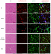
Figure 7. Engraftment of human iPSC-derived muscle progenitors into immune-compromised mice
Representative H&E images and immunostaining on TA sections of pre-injured-NSG mice injected with 1×10⁵ iPSCs at day 14 of differentiation. Muscles injected with BJ, 00409, or 05400 iPSC-derived cells stain positively for humanδ-Sarcoglycan protein(red). Fibers were counterstained with Laminin (green). No staining is observed in PBS injected mice or when 00409 fibroblast cells were transplanted. Because the area of human cell engraftment could not be specifically distinguished on Hematoxylin/Eos instained sections, which must be processed differently from sections for immunostaining, the H&E images shown do not represent the same muscle region as that shown in immunofluorescence images. Scale bars represent 100 μm, n=3 per sample. See also Figure S10.

Comment in

References

    1. Awaya T, Kato T, Mizuno Y, Chang H, Niwa A, Umeda K, Nakahata T, Heike T. Selective development of myogenic mesenchymal cells from human embryonic and induced pluripotent stem cells. PLoS One. 2012;7:e51638. - PMC - PubMed
    1. Barberi T, Bradbury M, Dincer Z, Panagiotakos G, Socci ND, Studer L. Derivation of engraftable skeletal myoblasts from human embryonic stem cells. Nat Med. 2007a;13:642–648. - PubMed
    1. Barberi T, Bradbury M, Dincer Z, Panagiotakos G, Socci ND, Studer L. Derivation of engraftable skeletal myoblasts from human embryonic stem cells. Nature Medicine. 2007b;13:642–648. - PubMed
    1. Brack AS, Bildsoe H, Hughes SM. Evidence that satellite cell decrement contributes to preferential decline in nuclear number from large fibres during murine age-related muscle atrophy. J Cell Sci. 2005;118:4813–4821. - PubMed
    1. Brack AS, Conboy IM, Conboy MJ, Shen J, Rando TA. A temporal switch from notch to Wnt signaling in muscle stem cells is necessary for normal adult myogenesis. Cell Stem Cell. 2008;2:50–59. - PubMed

Publication types

MeSH terms

LinkOut - more resources