. 2013 Nov 14;5(3):619-28.
doi: 10.1016/j.celrep.2013.10.011. Epub 2013 Nov 7.

SHARPIN regulates uropod detachment in migrating lymphocytes

Affiliations

SHARPIN regulates uropod detachment in migrating lymphocytes

Jeroen Pouwels et al. Cell Rep. .

Abstract

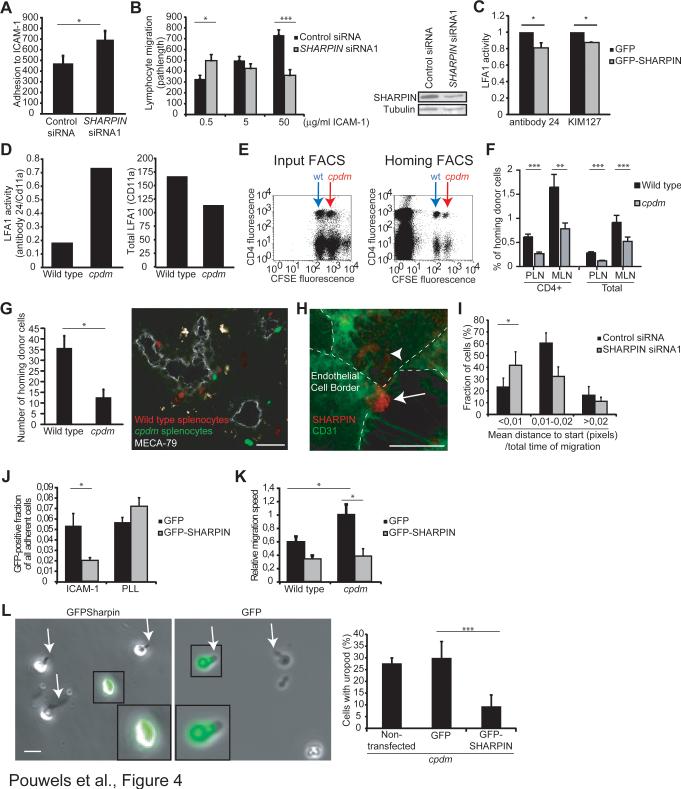
SHARPIN-deficient mice display a multiorgan chronic inflammatory phenotype suggestive of altered leukocyte migration. We therefore studied the role of SHARPIN in lymphocyte adhesion, polarization, and migration. We found that SHARPIN localizes to the trailing edges (uropods) of both mouse and human chemokine-activated lymphocytes migrating on intercellular adhesion molecule-1 (ICAM-1), which is one of the major endothelial ligands for migrating leukocytes. SHARPIN-deficient cells adhere better to ICAM-1 and show highly elongated tails when migrating. The increased tail lifetime in SHARPIN-deficient lymphocytes decreases the migration velocity. The adhesion, migration, and uropod defects in SHARPIN-deficient lymphocytes were rescued by reintroducing SHARPIN into the cells. Mechanistically, we show that SHARPIN interacts directly with lymphocyte-function-associated antigen-1 (LFA-1), a leukocyte counterreceptor for ICAM-1, and inhibits the expression of intermediate and high-affinity forms of LFA-1. Thus, SHARPIN controls lymphocyte migration by endogenously maintaining LFA-1 inactive to allow adjustable detachment of the uropods in polarized cells.


Figures

Figure 1
Figure 1. SHARPIN Regulates Uropod Detachment in Migrating Lymphocytes
(A) CXCL12-stimulated, wild-type and Sharpin-deficient (Sharpincpdm) mouse splenocytes migrating on recombinant mouse ICAM-1 (5 μg/ml) immunostained for SHARPIN and CD44. Representative micrographs and quantitation of mean fluorescence intensities (MFI) along the cell axis (mean±sem (sem in grey color); n >10 cells/staining; statistical comparisons between the 20 first frontal and 20 last posterior measurement points) are shown. Bars, 10 μm. (B, C) Adhesion (B) and spreading (C) of wild-type and Sharpincpdm splenocytes and thymocytes on ICAM-1 (5 μg/ml). (D, E) Migration of CXCL12-stimulated, wild-type and Sharpincpdm (D) splenocytes and (E) purified CD4+ T cells on different concentrations of ICAM-1. Representative cell tracks and cumulative data are shown. See also Video S1 and Video S2. (F) Phenotype of the uropods in cells migrating on high ICAM-1 concentration (50 μg/ml) was determined using image analyses. Extended uropod defines an uropod length > 33% of cell body diameter, polarity index is the ratio of the longest leading edge-to-uropod distance divided by the longest width of the cell and uropod lifetime is the time difference between uropod appearance and disappearance. Bars, 10 μm. (G) Expression of SHARPIN, CD44, F-actin and talin in purified, CXCL12-stimulated CD4+ wild-type and Sharpincpdm cells migrating on high ICAM-1 concentration (50 μg/ml). Representative micrographs and quantitations as in (A) (n = 5-8 cells/staining) are shown. Bars, 10 μm. Data are representative from or are the mean±sem of 3 (A-D, F) and 2 (E, G) independent experiments. In A, F and G the white arrows point to the uropods/tails. *p<0.05, **p<0.01, ***p<0.001 (Student's two-tailed t-test).
Figure 2
Figure 2. SHARPIN and Inactive LFA-1 Co-Localize in Uropods
(A, B) Expression of LFA-1 on isolated wild-type and Sharpincpdm thymocytes and total and CD4-positive splenocytes. Shown are (A) representative FACS plots, and (B) the specific mean fluorescence intensities (MFI) for CD11a. (C) Immunostainings of cytospin preparations of resting human PBMC with anti-SHARPIN and control Abs. DAPI was used to visualize nuclear morphology. An arrowhead points to a representative lymphocyte. Bars, 10 μm. (D) Immunostainings of freshly isolated, CXCL12-stimulated human PBMC migrating on recombinant human ICAM-1 (5 μg/ml) for SHARPIN (a non-polarized and polarized cell are shown). (E-F) Immunostainings of GFP-SHARPIN transfected, CXCL12-stimulated human PBMC migrating on ICAM for (E) CD44, and (F) talin. (G-I) Immunostainings of freshly isolated, CXCL12-stimulated human PBMC migrating on ICAM-1 for SHARPIN and, (G) CD11a (total), (H) extended LFA-1 (KIM127 epitope) and (I) activated LFA-1 (24 epitope). (J) Quantitations of mean fluorescence intensities (MFI) of the indicated stainings along the cell axis (mean±sem (sem in grey color); n > 4-8 cells/staining; statistical comparisons between the 20 first frontal and 20 last posterior measurement points) are shown. In (D-I) the white arrows point to the uropods, and the bars are 5 μm. Data in (A-B) are from 4-9 mice/genotype and in (C-I) representative of at least 3 independent experiments with different blood cell donors. *p<0.05, **p<0.01, ***p<0.001 (Student's two-tailed t-test). See also Figure S1.
Figure 3
Figure 3. SHARPIN Directly Binds to the Conserved Membrane Proximal Sequence of αL
(A-C) Co-immunoprecipitation of SHARPIN with LFA-1 in (A) HEK 293 cells overexpressing αL and β2 integrins and GFP or GFP-SHARPIN, (B) Jurkat cells (endogenous proteins) and (C) freshly isolated PBMC (endogenous proteins). (D) The integrin constructs used in the binding studies (ECD, extracellular domain; TMD, transmembrane domain and CYPD; cytoplasmic domain) and the sequences of the transmembrane and intracellular domains of αL, αM, αX, αD and β2 integrins. The conserved membrane proximal area in α-tails is highlighted in red. (E) Pull-down experiments with recombinant GST-SHARPIN and recombinant full-length LFA-1 and LFA-1 lacking the cytoplasmic tails. GST only is a loading control. (F) Interaction of different GST-SHARPIN with the full-length and tail-less LFA-1 heterodimers was determined using far western overlay assays. (G) Pull-down assays with synthetic biotinylated peptides corresponding to the cytoplasmic sequences of αL, αM, αD and β2 with lysates of GFP-SHARPIN transfected cells. αL cons, the conserved membrane proximal part of αL. Data are representative of at least 3 independent experiments. See also Figure S2.
Figure 4
Figure 4. Sharpin De-Activates LFA-1, Improves Lymphocyte Extravasation and Re-Expression of SHARPIN Rescues the Uropod and Migration Defects of SHARPIN-Deficient Cells
(A-B) Adhesion (A) and migration (B) of human PBMC transfected with SHARPIN siRNA or control siRNA on recombinant human ICAM-1, and the efficacy of knockdown on SHARPIN protein levels. (C) FACS analyses of the reporter mAb 24 and KIM127 cell surface binding to GFP-SHARPIN and control (GFP only) expressing, CXCL12-stimulated human PBMC. The results are normalized against total CD11a expression. (D) Cell surface expression of 24 reporter epitope (normalized to total αL expression; left panel) and total αL expression (MFI, right panel) on wild-type and Sharpincpdm MEFs expressing human LFA-1 was determined using FACS (a representative experiment). (E) Representative FACS plots of the input population and transferred cells isolated from mesenteric lymph nodes of wild-type recipient mice in the homing experiments after 2 h. Wild-type (indicated by blue arrow) and Sharpincpdm (indicated by red arrow) donor splenocytes are CFSE dim and bright, respectively, and the cells were co-stained with Alexa 647-labeled anti-CD4. (F) Percentages of wild-type and Sharpincpdm CD4-positive and total lymphocytes in the mesenteric (MLN) and peripheral (PLN) lymph nodes of wild-type recipient mice after a 2 h recirculation period (n = 7 mice/genotype). (G) Microscopic analyses of homing of wild-type (red) and Sharpincpdm (green) cells to PLN after a 30 min recirculation. MECA-79 positive high endothelial venules are shown in grey. A representative micrograph and pooled data (n = 7 recipient mice; 187 homed wild-type cells) are shown. Bar, 50 μm. (H, I) Transmigration of human PBMC transfected with SHARPIN siRNA or control siRNA through an endothelial monolayer in in vitro flow assays. (H) Expression of SHARPIN in a PBMC transmigrating through CD31+ endothelial monolayer (endothelial borders are shown by a dashed line, and the arrow indicates the leukocyte uropod, and the arrowhead the PBMC cell body below the endothelium). (I) Net displacement of PBMC after transmigration (n = 40 control siRNA and n = 45 SHARPIN siRNA transfected cells from 3 independent experiments with different blood and endothelial cell donors). See also Video S3. (J) Adhesion assays determining the binding of wild-type and Sharpincpdm splenocytes expressing GFP-SHARPIN or GFP alone to ICAM-1 and poly-L-lysine (PLL, a nonintegrin binding ligand). (K) Migration velocity of wild-type and Sharpincpdm splenocytes expressing GFPSHARPIN or GFP alone on ICAM-1. (L) Uropod formation on Sharpincpdm cells expressing SHARPIN-GFP or GFP alone after CXCL12-stimulation and migration on ICAM-1 (50 μg/ml). Data are the mean±sem of 3 (A, I), 2 (B, G, J-L), 2-5 (C) independent experiments. *p<0.05, **p<0.01, ***p<0.001 (Student's two-tailed t-test). See also Figure S3.

References

    1. Abram CL, Lowell CA. The ins and outs of leukocyte integrin signaling. Annu Rev Immunol. 2009;27:339–362. - PMC - PubMed
    1. Bolomini-Vittori M, Montresor A, Giagulli C, Staunton D, Rossi B, Martinello M, Constantin G, Laudanna C. Regulation of conformer-specific activation of the integrin LFA-1 by a chemokine-triggered Rho signaling module. Nat Immunol. 2009;10:185–194. - PubMed
    1. Constantin G, Majeed M, Giagulli C, Piccio L, Kim JY, Butcher EC, Laudanna C. Chemokines trigger immediate beta2 integrin affinity and mobility changes: differential regulation and roles in lymphocyte arrest under flow. Immunity. 2000;13:759–769. - PubMed
    1. Dransfield I, Hogg N. Regulated expression of Mg2+ binding epitope on leukocyte integrin a subunits. EMBO J. 1989;8:3759–3765. - PMC - PubMed
    1. Franco SJ, Rodgers MA, Perrin BJ, Han J, Bennin DA, Critchley DR, Huttenlocher A. Calpain-mediated proteolysis of talin regulates adhesion dynamics. Nat Cell Biol. 2004;6:977–983. - PubMed

Publication types

Substances