. 2013 Nov 14;5(3):826-38.
doi: 10.1016/j.celrep.2013.10.008. Epub 2013 Nov 7.

DUB-resistant ubiquitin to survey ubiquitination switches in mammalian cells

Affiliations

DUB-resistant ubiquitin to survey ubiquitination switches in mammalian cells

Miklós Békés et al. Cell Rep. .

Abstract

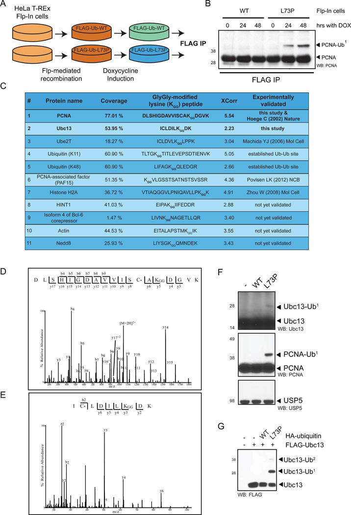
The ubiquitin-modification status of proteins in cells is highly dynamic and maintained by specific ligation machineries (E3 ligases) that tag proteins with ubiquitin or by deubiquitinating enzymes (DUBs) that remove the ubiquitin tag. The development of tools that offset this balance is critical in characterizing signaling pathways that utilize such ubiquitination switches. Herein, we generated a DUB-resistant ubiquitin mutant that is recalcitrant to cleavage by various families of DUBs both in vitro and in mammalian cells. As a proof-of-principle experiment, ectopic expression of the uncleavable ubiquitin stabilized monoubiquitinated PCNA in the absence of DNA damage and also revealed a defect in the clearance of the DNA damage response at unprotected telomeres. Importantly, a proteomic survey using the uncleavable ubiquitin identified ubiquitinated substrates, validating the DUB-resistant ubiquitin expression system as a valuable tool for interrogating cell signaling pathways.


Conflict of interest statement

Conflicts of Interest

B.B. and F.M. are employees of Boston Biochem, Inc. The rest of the authors declare no conflicts of interest.

Figures

Figure 1
Figure 1. UbL73P is a pan-DUB DUB-resistant ubiquitin mutant in vitro
(A) Surface representation of ubiquitin (PDB: 1UBQ) detailing its C-terminus. The DUB cleavage site (P4-P1, aa 72–76 of ubiquitin) is shown in sticks (in black), with Leu73 shown in green. The Ile44 hydrophobic patch is colored brown on the surface, while the Ile36 hydrophobic patch is colored pink. The image was generated using PyMol. (B) Schematic representation of Smt3-tagged linear di-ubiquitin. Ubiquitin L73P is not cleaved by DUBs neither as a peptide or an iso-peptide bond. Wild-type and L73P Smt3-linear-di-Ub, before (C) and after (D) Ulp1-cleavage and final purification, was cleaved with a serial dilution of USP2CD. (E) Untagged mono-ubiquitins were used to make K63-linked di-ubiquitins using Ubc13/Uev1a, then subjected to cleavage by USP2CD. (F) Recombinant USP1/UAF1 complex was used to cleave wild-type and L73P (NH) tetra-ubiquitin chains in vitro. (G) Linkage-specific tetra-ubiquitin chains (K48-linked, left panel, or K63-linked, right panel) made using either wild-type or L73P ubiquitin (non-hydrolyzable (NH) chains) were cleaved with an assortment of USP-family DUBs. (H) DUBs representing the USP-family (USP2CD), the OTU-domain family (Otubain-1) and the JAMM-metallo-DUBs (AMSH) were used cleave wild-type and NH K63-linked tetra-ubiquitin chains. Otubain-1 serves as a negative control for K63. (I) Otubain-1 and USP2CD were used to cleave K48-linked di-ubiquitin. (J) USP2CD was used to cleave K11-linked di-ubiquitin. Dotted lines indicate cropping within the same gel. Assays were carried out at 37°C for 1 hour and analyzed by SDS-PAGE and coomassie-staining. See also Figure S1.
Figure 2
Figure 2. UbL73P is conjugated to substrates in vitro and in vivo
(A) E1 charging assay with GST-tagged human Ube1 and Uba6 using wild-type and L73P ubiquitin. (B) E2 charging assay using human GST-tagged Ube1 and Uba6 as the E1 and human UbcH7 as the E2, using wild-type and L73P ubiquitin (F). E1 and E2 charging reactions were carried out at 37°C in the absence of DTT and analyzed by non-reducing SDS-PAGE and SYPRO-staining. (C) and (D) p53-ubiquitination reactions with wild-type and L73P ubiquitin using either a RING-finger E3 ligase, Mdm2 (C) or a HECT-domain E3 ligase, E6AP (D). Ubiquitination reactions with the E3 ligases were analyzed by denaturing SDS-PAGE and western blotting with an anti-p53 antibody. Dotted lines indicate cropped images from the same gel. (E) Mdm2-generated ubiquitin chains on p53 using WT or L73P ubiquitin were cleaved with USP2CD following ubiquitination reactions. (F) Expression of HA-tagged wild-type and L73P ubiquitin in U2OS cells results in stabilization of Ub-conjugates by L73P, such as PCNA-Ub. See also Figure S2.
Figure 3
Figure 3. Dynamic PCNA mono-ubiquitination revealed by UbL73P
(A) HA-Ub constructs were expressed in HEK293 cells, lysed under non-denaturing conditions and the lysates were treated with a serial dilution of USP2CD. (B) HA-Ub constructs were expressed in U2OS cells and lysed under denaturing conditions. (C) PCNA with a K164R mutation does not support mono-ubiquitination by UbL73P. HA-PCNA and myc-ubiquitin constructs were co-expressed in U2OS cells and lysed under denaturing conditions. (D) UbL73P is conjugated in a Rad18-dependent manner in vivo. U2OS cells were treated with siRNAs for 24 hours, then transfected with HA-ubiquitin for another 48 hours when cell lysates were prepared under denaturing conditions. (E) UbL73P-stabilized PCNA-Ub recruits polymerase eta in the absence of DNA damage. HA-tagged Ubs were co-expressed with GFP-tagged Pol eta in U2OS cells, fixed in methanol, mounted with DAPI and analyzed by immunofluorescence microscopy using anti-GFP antibodies for nuclear GFP-ETA foci. The inlets show representative images and western blots for the cell lysates analyzed. Error bars represent standard deviation of the mean, n=3. (F) Dynamic PCNA mono-ubiquitination revealed by UbL73P. Under normal circumstance in S-phase (top panel) the E3 ligase Rad18 mono-ubiquitinates PCNA, which is counter-balanced by USP1, the DUB for PCNA. When a conjugatable, but not deconjugatable ubiquitin (L73P, bottom panel) is expressed in cells, the USP1-arm of the dynamic PCNA monoubiquitation cycle is poisoned, leading to the stabilization of PCNA-Ub. See also Figure S3.
Figure 4
Figure 4. Ubiquitin-conjugate stabilization reveals a telomeric DNA damage response phenotype
RAP80-mediated pull-down of UbL73P-conjugates. (A) and (B) HA-ubiquitin constructs were expressed in HEK293 cells, lysed under non-denaturing conditions and Ub-conjugates were pulled-down with RAP80-conjugated beads, then analyzed by SDS-PAGE and western blotting for the indicated antibodies. (C) TRF2F/F; Rosa26 CRE-ER MEFs infected with the indicated constructs were treated with 4-hydroxytamoxifen (+OHT) and stained for 53BP1 (red), telomere DNA (green), and DAPI (blue). (D) Quantification of cells with less than 10 53BP1 foci (<10) or more than 30 53BP1 foci (>30) per nucleus at the indicated time points following OHT treatment. (E) Metaphase spreads were harvested from TRF2F/F; Rosa26 CRE-ER MEFs seven days following OHT treatment. Cells were infected with either HA-Ub or HA-UbL73P and stained for telomere DNA (green) and DAPI (red). Percentages of chromosomes with fusions are indicated. Bar graph in the lower panel indicates the percentage of chromosome fusions in MEFs infected with the indicated constructs and either treated with tamoxifen (+OHT) or untreated (−OHT). See also Figure S4.
Figure 5
Figure 5. UbL73P reveals stabilized ubiquitination of Ubc13 and other targets
(A) Schematics for the generation of HeLa Flp-in cells stably expressing ubiquitin constructs. (B) Anti-FLAG immune-precipitation of Flp-in cells treated for the indicated times with doxycycline to induce transgene expression, probed with anti-PCNA antibody. (C) Selected list of proteins conjugated by UbL73P and the peptides containing the Ub-modified Lys residues. (D) MS/MS spectrum of the doubly charged ion of the peptide (DLSHIGDAVVISCAKGGDGVK carboxymethylated on the Cys (*) residue and carrying a GlyGly modification on the first lysine residue) for Lys164 of PCNA, (E) MS/MS spectrum of the triply charged ion of the peptide (ICLDILKGGDK carboxymethylated on the Cys (*) residue and carrying a GlyGly modification on the first lysine residue) for Lys92 of Ubc13. Observed peptide bond cleavages are indicated in the sequences. The corresponding theoretical N-terminal (b-type ion) and C-terminal (y-type ions) ion series for the observed fragment ions are shown above and below the sequence, respectively. Neutral loss of water from y- and b- type ions are not indicated in the spectra. (F) HA-Ub constructs were expressed in U2OS cells, lysed under non-denaturing conditions and probed with the indicated antibodies. (G) FLAG-Ubc13 and HA-Ub constructs were co-expressed in U2OS cells, lysed under denaturing conditions and probed with anti-FLAG antibody. See also Figure S4 and Table S1.

References

    1. Bekes M, Prudden J, Srikumar T, Raught B, Boddy MN, Salvesen GS. The dynamics and mechanism of sumo chain deconjugation by SUMO-specific proteases. J Biol Chem. 2011;286:10238–10247. - PMC - PubMed
    1. Bohren KM, Gabbay KH, Owerbach D. Affinity chromatography of native SUMO proteins using His-tagged recombinant UBC9 bound to Co2+-charged talon resin. Protein Expr Purif. 2007;54:289–294. - PubMed
    1. Celli GB, de Lange T. DNA processing is not required for ATM-mediated telomere damage response after TRF2 deletion. Nat Cell Biol. 2005;7:712–718. - PubMed
    1. Cesare AJ, Hayashi MT, Crabbe L, Karlseder J. The telomere deprotection response is functionally distinct from the genomic DNA damage response. Mol Cell. 2013;51:141–155. - PMC - PubMed
    1. Cotto-Rios XM, Bekes M, Chapman J, Ueberheide B, Huang TT. Deubiquitinases as a signaling target of oxidative stress. Cell reports. 2012;2:1475–1484. - PMC - PubMed

Publication types

Substances