. 2013 Nov 27;5(4):909-17.
doi: 10.1016/j.celrep.2013.10.012. Epub 2013 Nov 7.

Changes in cellular mRNA stability, splicing, and polyadenylation through HuR protein sequestration by a cytoplasmic RNA virus

Affiliations

Changes in cellular mRNA stability, splicing, and polyadenylation through HuR protein sequestration by a cytoplasmic RNA virus

Michael D Barnhart et al. Cell Rep. .

Abstract

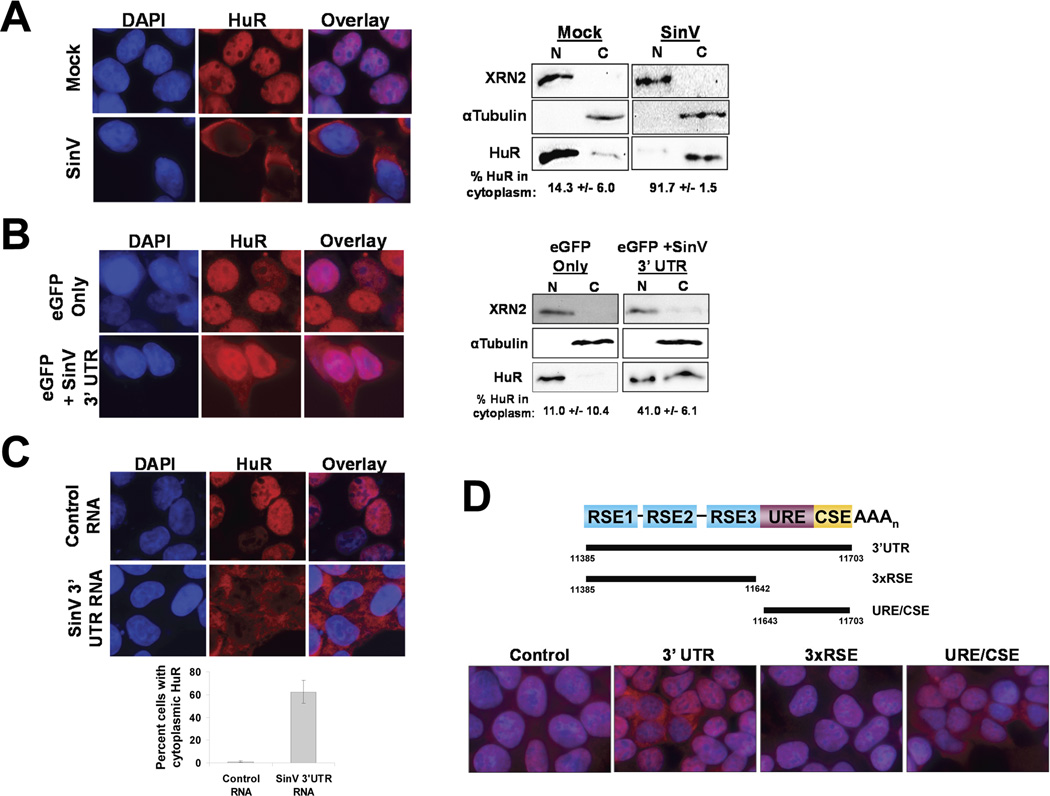
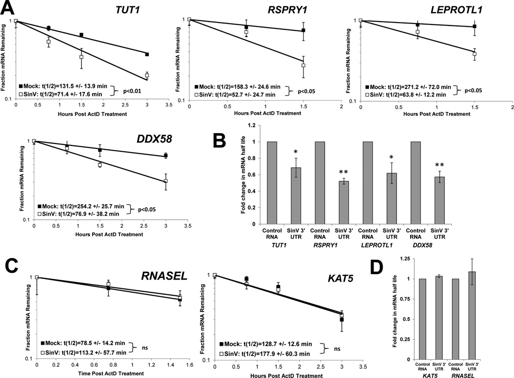
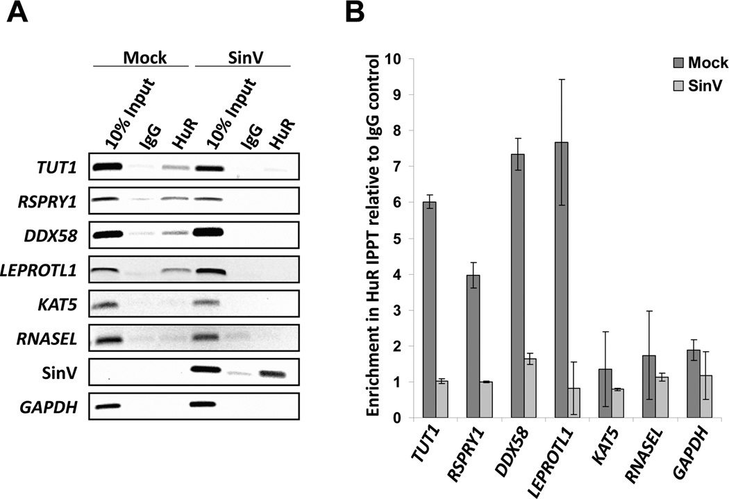
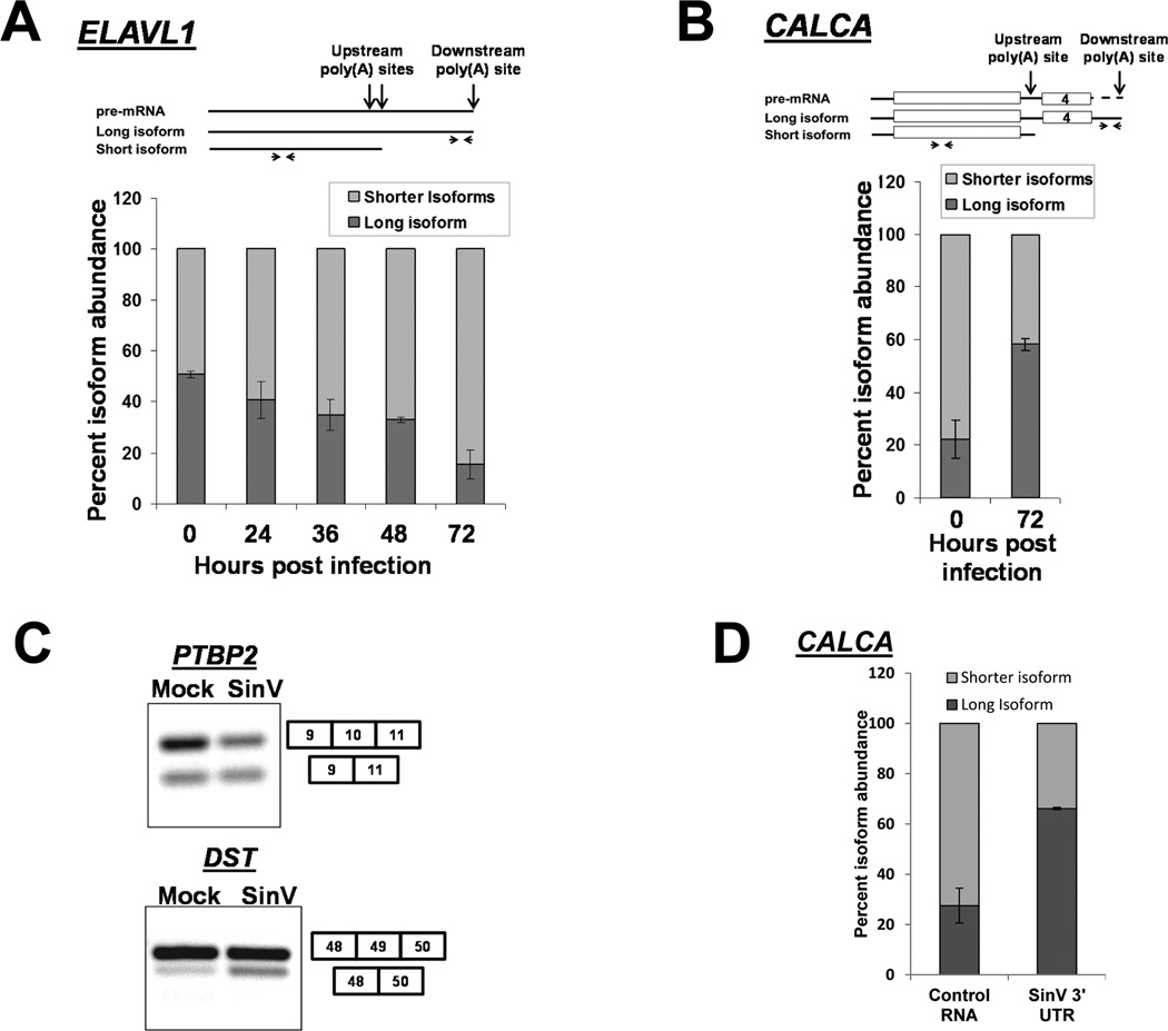
The impact of RNA viruses on the posttranscriptional regulation of cellular gene expression is unclear. Sindbis virus causes a dramatic relocalization of the cellular HuR protein from the nucleus to the cytoplasm in infected cells. This is to the result of the expression of large amounts of viral RNAs that contain high-affinity HuR binding sites in their 3' UTRs effectively serving as a sponge for the HuR protein. Sequestration of HuR by Sindbis virus is associated with destabilization of cellular mRNAs that normally bind HuR and rely on it to regulate their expression. Furthermore, significant changes can be observed in nuclear alternative polyadenylation and splicing events on cellular pre-mRNAs as a result of sequestration of HuR protein by the 3' UTR of transcripts of this cytoplasmic RNA virus. These studies suggest a molecular mechanism of virus-host interaction that probably has a significant impact on virus replication, cytopathology, and pathogenesis.


Figures

Figure 1
Figure 1. The SinV 3’ UTR is sufficient to induce the relocalization of HuR protein from the nucleus to the cytoplasm
A. 293T cells were mock-treated or infected with SinV. At 24 hrs post treatment, cells were analyzed for HuR protein localization by immunofluorescence (left panel) or by subcellular fractionation and western blotting (right panel) using antibodies specific for the indicated proteins. DAPI was used to identify the nuclear compartment. Panel B. Cells were transfected with plasmids encoding an eGFP reporter bearing either the default 3’ UTR (eGFP only) or the SinV 3’ UTR (eGFP + SinV 3’ UTR) and analyzed for HuR protein subcellular localization by immunofluorescence (left panel) or by subcellular fractionation and western blotting (right panel). Panel C: 293T cells were transfected with a control RNA or with a SinV 3’ UTR RNA. At 6 hrs post transfection, cells were analyzed for HuR protein subcellular localization by immunofluorescence. Quantification is shown with standard deviations. Panel D: 293T cells were transfected with a control RNA or with the indicated fragment of the SinV 3’ UTR (diagrammed at the top of the panel). At 6 hrs post transfection, cells were analyzed for HuR protein subcellular localization by immunofluorescence.
Figure 2
Figure 2. SinV infection influences the stability of some but not all cellular mRNAs
At 24 hpi with SinV, 293T cells were treated with actinomycin D and the relative levels of the indicated mRNAs were assessed at the designated time points following shut off of transcription using qRT-PCR to determine mRNA half-lives. Panel A depicts mRNAs that were destabilized during SinV infection while panel C contains mRNAs whose stability was not affected. Representative decay curves are shown with standard deviation of experimental measurements and the average half-lives are reported with standard deviations from three independent experiments. In Panels B and D, 293T cells were transfected with equimolar amounts of either a control RNA (GemA60) or the SinV 3’UTR RNA. At 4.5 hrs post transfection, cells were treated with actinomycin D and the relative levels of the indicated mRNAs were assessed by qRT-PCR. Average fold change in half-lives are reported with standard deviations from two independent experiments. * p<0.05; ** p<0.01.
Figure 3
Figure 3. SinV infection significantly reduced the association of HuR with cellular mRNAs
293T cells were either mock-infected or infected with SinV. At 24 hrs post treatment, HuR protein-RNA complexes were isolated by immunoprecipitation using HuR-specific antibodies or a control normal mouse IgG. Co-precipitating mRNAs were analyzed by RT-PCR (panel A) or qRT-PCR (panel B). The two panels depict results from independent infections.
Figure 4
Figure 4. SinV infection influences alternative polyadenylation and splicing of cellular pre-mRNAs
293T cells were infected with SinV for the indicated times. Short or long isoforms of the ELAVL1 (HuR) mRNA formed by alternative polyadenylation (Panel A) or the indicated isoforms of CALCA (calcitonin) were quantified by qRT-PCR using the primers illustrated at the top of the panels. Note that the level of ‘shorter isoforms’ was determined by subtraction of the amount of the longer isoform from the level of total RNA detected by the upstream primer. Quantification is shown with standard deviations calculated from three independent experiments. Panel C. Total RNA was isolated 24 hpi, probed for the presence of the splicing isoforms of the indicated genes by RT-PCR and analyzed on a 2% agarose gel. Panel D: 293T cells were transfected with equimolar amounts of either a control RNA (GemA60) or the SinV 3’UTR RNA. Total RNA was isolated at 6 hours post transfection and analyzed for CALCA isoforms as described in Panel B above. Quantification is shown with standard deviations calculated from two independent experiments.

References

    1. Abdelmohsen K, Gorospe M. Posttranscriptional regulation of cancer traits by HuR. Wiley Interdiscip. Rev. RNA. 2010;1:214–229. - PMC - PubMed
    1. Atasheva S, Gorchakov R, English R, Frolov I, Frolova E. Development of Sindbis viruses encoding nsP2/GFP chimeric proteins and their application for studying nsP2 functioning. J. Virol. 2007;81:5046–5057. - PMC - PubMed
    1. Benoit RM, Meisner NC, Kallen J, Graff P, Hemmig R, Cèbe R, Ostermeier C, Widmer H, Auer M. The x-ray crystal structure of the first RNA recognition motif and site-directed mutagenesis suggest a possible HuR redox sensing mechanism. J. Mol. Biol. 2010;397:1231–1244. - PubMed
    1. Bevilacqua A, Ghisolfi L, Franzi S, Maresca G, Gherzi R, Capaccioli S, Nicolin A, Canti G. Stabilization of cellular mRNAs and up-regulation of proteins by oligoribonucleotides homologous to the Bcl2 adenine-uridine rich element motif. Mol. Pharmacol. 2007;71:531–538. - PubMed
    1. Bolognani F, Gallani AI, Sokol L, Baskin DS, Meisner-Kober N. mRNA stability alterations mediated by HuR are necessary to sustain the fast growth of glioma cells. J. Neurooncol. 2012;106:531–542. - PubMed

Publication types

MeSH terms