Canine Mammary Cancer Stem Cells are Radio- and Chemo- Resistant and Exhibit an Epithelial-Mesenchymal Transition Phenotype
- PMID: 24212780
- PMCID: PMC3757388
- DOI: 10.3390/cancers3021744
Canine Mammary Cancer Stem Cells are Radio- and Chemo- Resistant and Exhibit an Epithelial-Mesenchymal Transition Phenotype
Erratum in
-
Correction: Pang et al. Canine Mammary Cancer Stem Cells Are Radio- and Chemo- Resistant and Exhibit an Epithelial-Mesenchymal Transition Phenotype. Cancers 2011, 3, 1744-1762.Cancers (Basel). 2022 Aug 31;14(17):4242. doi: 10.3390/cancers14174242. Cancers (Basel). 2022. PMID: 36077879 Free PMC article.
Abstract
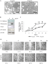
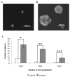
Canine mammary carcinoma is the most common cancer among female dogs and is often fatal due to the development of distant metastases. In humans, solid tumors are made up of heterogeneous cell populations, which perform different roles in the tumor economy. A small subset of tumor cells can hold or acquire stem cell characteristics, enabling them to drive tumor growth, recurrence and metastasis. In veterinary medicine, the molecular drivers of canine mammary carcinoma are as yet undefined. Here we report that putative cancer stem cells (CSCs) can be isolated form a canine mammary carcinoma cell line, REM134. We show that these cells have an increased ability to form tumorspheres, a characteristic of stem cells, and that they express embryonic stem cell markers associated with pluripotency. Moreover, canine CSCs are relatively resistant to the cytotoxic effects of common chemotherapeutic drugs and ionizing radiation, indicating that failure of clinical therapy to eradicate canine mammary cancer may be due to the survival of CSCs. The epithelial to mesenchymal transition (EMT) has been associated with cancer invasion, metastasis, and the acquisition of stem cell characteristics. Our results show that canine CSCs predominantly express mesenchymal markers and are more invasive than parental cells, indicating that these cells have a mesenchymal phenotype. Furthermore, we show that canine mammary cancer cells can be induced to undergo EMT by TGFβ and that these cells have an increased ability to form tumorspheres. Our findings indicate that EMT induction can enrich for cells with CSC properties, and provide further insight into canine CSC biology.
Figures


References
-
- Benjamin S.A., Lee A.C., Saunders W.J. Classification and behavior of canine mammary epithelial neoplasms based on life-span observations in beagles. Vet. Pathol. 1999;36:423–436. - PubMed
-
- Fidler I.J., Brodey R.S. The biological behavior of canine mammary neoplasms. J. Am. Vet. Med. Assoc. 1967;151:1311–1318. - PubMed
-
- Priester W.A., Mantel N. Occurrence of tumors in domestic animals. Data from 12 united states and canadian colleges of veterinary medicine. J. Natl. Cancer Inst. 1971;47:1333–1344. - PubMed
-
- Cohen D., Reif J.S., Brodey R.S., Keiser H. Epidemiological analysis of the most prevalent sites and types of canine neoplasia observed in a veterinary hospital. Cancer Res. 1974;34:2859–2868. - PubMed
-
- Schneider R., Dorn C.R., Taylor D.O. Factors influencing canine mammary cancer development and postsurgical survival. J. Natl. Cancer Inst. 1969;43:1249–1261. - PubMed
LinkOut - more resources
Full Text Sources

