. 2012 Feb 22;4(1):218-43.
doi: 10.3390/cancers4010218.

Tumor microenvironment in the brain

Affiliations

Tumor microenvironment in the brain

Mihaela Lorger. Cancers (Basel). .

Abstract

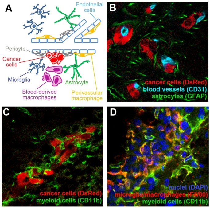
In addition to malignant cancer cells, tumors contain a variety of different stromal cells that constitute the tumor microenvironment. Some of these cell types provide crucial support for tumor growth, while others have been suggested to actually inhibit tumor progression. The composition of tumor microenvironment varies depending on the tumor site. The brain in particular consists of numerous specialized cell types such as microglia, astrocytes, and brain endothelial cells. In addition to these brain-resident cells, primary and metastatic brain tumors have also been shown to be infiltrated by different populations of bone marrow-derived cells. The role of different cell types that constitute tumor microenvironment in the progression of brain malignancies is only poorly understood. Tumor microenvironment has been shown to be a promising therapeutic target and diagnostic marker in extracranial malignancies. A better understanding of tumor microenvironment in the brain would therefore be expected to contribute to the development of improved therapies for brain tumors that are urgently required due to a poor availability of treatments for these malignancies. This review summarizes some of the known interactions between brain tumors and different stromal cells, and also discusses potential therapeutic approaches within this context.


Figures

Figure 1
Figure 1
Tumor microenvironment in the brain consists of different cell types. (A) Schematic representation of some major cell types that constitute tumor microenvironment in the brain. (B) Mouse breast carcinoma cell line EO771 that stably expresses red fluorescent protein (DsRed; red) metastasized to the brain upon the administration of cancer cells into the internal carotid artery. Extravasated cancer cells remained associated with the blood vessels (light blue). The presence of cancer cells induced a strong activation of astrocytes, as demonstrated by their increased expression of GFAP (green). (C) Small metastatic lesions of EO771 cells (red) are surrounded by CD11b+ cells of myeloid origin with different morphologies (green). (D) A larger metastatic lesion established by EO771 cells (nuclear staining in blue) is infiltrated by CD11b+ myeloid cells (green). Some of these cells are macrophages/microglia as demonstrated by their positive staining for F4/80 (red).

References

    1. Al-Shamy G., Sawaya R. Management of brain metastases: The indispensable role of surgery. J. Neurooncol. 2009;92:275–282. doi: 10.1007/s11060-009-9839-y. - DOI - PubMed
    1. DeAngelis L.M. Treatment of brain metastasis. J. Support. Oncol. 2008;6:87–88. - PubMed
    1. Gavrilovic I.T., Posner J.B. Brain metastases: Epidemiology and pathophysiology. J. Neurooncol. 2005;75:5–14. doi: 10.1007/s11060-004-8093-6. - DOI - PubMed
    1. Gerrard G.E., Franks K.N. Overview of the diagnosis and management of brain, spine, and meningeal metastases. J. Neurol. Neurosurg. Psychiatr. 2004;75:ii37–ii42. doi: 10.1136/jnnp.2004.040493. - DOI - PMC - PubMed
    1. Nikiforova M.N., Hamilton R.L. Molecular diagnostics of gliomas. Arch. Pathol. Lab. Med. 2011;135:558–568. - PubMed

LinkOut - more resources