. 2013 Dec 12;504(7479):287-90.
doi: 10.1038/nature12682. Epub 2013 Nov 10.

A chain mechanism for flagellum growth

Affiliations

A chain mechanism for flagellum growth

Lewis D B Evans et al. Nature. .

Abstract

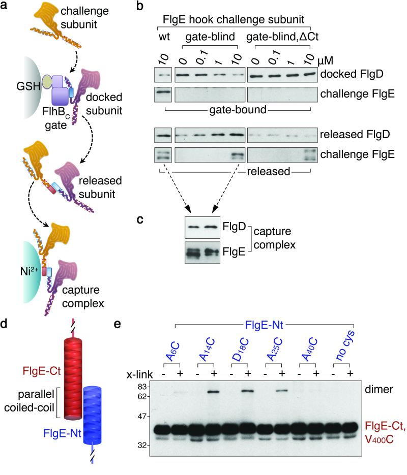
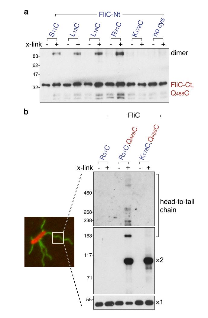
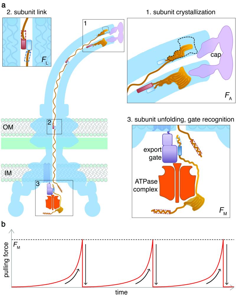
Bacteria swim by means of long flagella extending from the cell surface. These are assembled from thousands of protein subunits translocated across the cell membrane by an export machinery at the base of each flagellum. Unfolded subunits then transit through a narrow channel at the core of the growing flagellum to the tip, where they crystallize into the nascent structure. As the flagellum lengthens outside the cell, the rate of flagellum growth does not change. The mystery is how subunit transit is maintained at a constant rate without a discernible energy source in the channel of the external flagellum. We present evidence for a simple physical mechanism for flagellum growth that harnesses the entropic force of the unfolded subunits themselves. We show that a subunit docked at the export machinery can be captured by a free subunit through head-to-tail linkage of juxtaposed amino (N)- and carboxy (C)-terminal helices. We propose that sequential rounds of linkage would generate a multisubunit chain that pulls successive subunits into and through the channel to the flagellum tip, and by isolating filaments growing on bacterial cells we reveal the predicted chain of head-to-tail linked subunits in the transit channel of flagella. Thermodynamic analysis confirms that links in the subunit chain can withstand the pulling force generated by rounds of subunit crystallization at the flagellum tip, and polymer theory predicts that as the N terminus of each unfolded subunit crystallizes, the entropic force at the subunit C terminus would increase, rapidly overcoming the threshold required to pull the next subunit from the export machinery. This pulling force would adjust automatically over the increasing length of the growing flagellum, maintaining a constant rate of subunit delivery to the tip.


Figures

Figure 1
Figure 1. Free subunits capture subunits docked at the export gate via head-to-tail linkage of terminal helices
a, Capture of FlgD subunits docked at glutathione (GSH) bead-immobilised GSTFlhBC export gate component by free challenge subunits. Released capture complexes were collected by Ni2+ affinity chromatography. b, Docked FlgD subunit was challenged with increasing concentrations of free FlgE hook subunit, either wildtype (wt), unable to bind the export gate (gate-blind; Extended Data Fig. 3) or gate-blind and lacking C-terminal residues 359-403 (gate-blind, ΔCt). FlgE subunit lacking the C-terminus was attenuated for export (Extended Data Fig. 4e). c, Released capture complexes generated in b, (dashed arrows) were isolated confirming that captured FlgD is linked to challenge subunit. d, Juxtaposed carboxyl (FlgE-Ct) and amino (FlgE-Nt) terminal helices of sequential subunits adopt a parallel coiled-coil. e, Subunit pairs linked head-to-tail (dimer) using in vitro site-specific cysteine-cysteine cross-links. FlgE and variants containing unique cysteines (A6C, A14C, D18C, A25C or A40C; Extended Data Table 2) within and adjacent to the N-terminal helix predicted to generate the coiled-coil were incubated, with (+) or without (−) BMOE cross-linker (x-link) and a FlgE variant (V400C) containing a unique cysteine within the C-terminal helix. FlgE derivatives lacked either C- or N-termini (FlgE-Nt, FlgE-Ct) to preclude self-interaction. No dimers were detected for subunits without engineered cysteines (Extended Data Fig. 5e). All experiments were carried out at least three times and were biological replicates.
Figure 2
Figure 2. Head-to-tail linkage of flagellin subunits assembles a chain in the flagellum growing on the bacterial cell surface
a, Flagellin pairs linked head-to-tail (dimer) using in vitro site-specific cysteine-cysteine cross-links. Flagellin (FliC-Nt, FliC-Ct) and its variants containing unique cysteines (S11C, L13C, L18C, R31C or K178C) within and adjacent to the N-terminal helix predicted to generate a coiled-coil were incubated, with (+) or without (−) BMOE cross-linker (x-link) and a FliC variant containing a unique cysteine (Q488C) within the C-terminal helix. No complexes were detected between subunits without engineered cysteines (Extended Data Fig. 6a) b, Trapping of flagellin linked head-to-tail in chains within flagella (labelled green) growing on Salmonella cells (labelled red) using in vivo site-specific cysteine-cysteine cross-linking. Cells expressing recombinant full-length flagellin containing engineered cysteines (right panels; left lane, negative control R31C; centre lane, R31C and Q488C predicted to trap chain; right lane K178C and Q488C negative control) were incubated with (+) or without (−) BMOE cross-linker (x-link). Flagellar filaments were then isolated, depolymerised and resolved (immunoblot, panel exposure times decrease from top to bottom) to reveal monomer (x1), dimer (x2) and higher order head-to-tail chains of flagellin. We estimate between 6-10 linked subunits in the largest species. Dimers are predicted to form additionally by cross-linking between assembled flagellin C-terminal cysteines (Q488C) that line the channel and subunits in transit, so dimers are also seen in the controls (Extended Data Fig. 6b, c). All experiments were carried out at least three times and were biological replicates.
Figure 3
Figure 3. An entropic chain mechanism for flagellum growth outside the cell
a, 1. Subunit crystallization beneath the cap foldase provides a strong anchor (force to break anchor, FA; Supplementary Information 2). 2. Sequential subunits are linked (force to break link, FL) head-to-tail in a chain by juxtaposed terminal helices forming parallel coiled-coils. 3. Subunits docked at the export ATPase are unfolded and docked by the export machinery. The N-terminal helix of the docked subunit is then captured (force to break docking FM) into the subunit chain by the free C-terminal helix of an exiting subunit in the flagellar channel. b, Successive rounds of subunit capture from the export machinery. As an unfolded subunit crystallizes, the pulling force at its free end increases to reach the FM threshold (dashed line) whereupon the next subunit is captured from the export machinery. The pulling force then drops rapidly as the new unfolded subunit enters the channel. This process repeats for each subunit captured into the chain.

References

    1. Akeda Y, Galán JE. Chaperone release and unfolding of substrates in type III secretion. Nature. 2005;437:911–915. - PubMed
    1. Chevance FF, Hughes KT. Coordinating assembly of a bacterial macromolecular machine. Nature Rev. Microbiol. 2008;6:455–465. - PMC - PubMed
    1. Abrusci P, et al. Architecture of the major component of the type III secretion system export apparatus. Nature Struct. Mol. Biol. 2013;20:99–104. - PMC - PubMed
    1. Turner L, Stern AS, Berg HC. Growth of flagellar filaments of Escherichia coli is independent of filament length. J. Bacteriol. 2012;194:2437–2442. - PMC - PubMed
    1. Galán JE. Energizing type III secretion machines: what is the fuel? Nature Struct. Mol. Biol. 2008;15:127–128. - PMC - PubMed

Publication types

Substances