. 2014 Jan;306(2):L162-9.
doi: 10.1152/ajplung.00095.2013. Epub 2013 Nov 8.

Deletion of airway cilia results in noninflammatory bronchiectasis and hyperreactive airways

Affiliations

Deletion of airway cilia results in noninflammatory bronchiectasis and hyperreactive airways

Sandra K Gilley et al. Am J Physiol Lung Cell Mol Physiol. 2014 Jan.

Abstract

The mechanisms for the development of bronchiectasis and airway hyperreactivity have not been fully elucidated. Although genetic, acquired diseases and environmental influences may play a role, it is also possible that motile cilia can influence this disease process. We hypothesized that deletion of a key intraflagellar transport molecule, IFT88, in mature mice causes loss of cilia, resulting in airway remodeling. Airway cilia were deleted by knockout of IFT88, and airway remodeling and pulmonary function were evaluated. In IFT88(-) mice there was a substantial loss of airway cilia on respiratory epithelium. Three months after the deletion of cilia, there was clear evidence for bronchial remodeling that was not associated with inflammation or apparent defects in mucus clearance. There was evidence for airway epithelial cell hypertrophy and hyperplasia. IFT88(-) mice exhibited increased airway reactivity to a methacholine challenge and decreased ciliary beat frequency in the few remaining cells that possessed cilia. With deletion of respiratory cilia there was a marked increase in the number of club cells as seen by scanning electron microscopy. We suggest that airway remodeling may be exacerbated by the presence of club cells, since these cells are involved in airway repair. Club cells may be prevented from differentiating into respiratory epithelial cells because of a lack of IFT88 protein that is necessary to form a single nonmotile cilium. This monocilium is a prerequisite for these progenitor cells to transition into respiratory epithelial cells. In conclusion, motile cilia may play an important role in controlling airway structure and function.

Keywords: bronchiectasis; hyperreactivity; lung; respiratory.


Figures
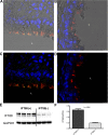
Fig. 1.
Fig. 1.
Representative immunofluorescence (IF) image of lung from an IFT88+ mouse shows ciliated respiratory epithelial cells of a large airway labeled with anti-acetylated α-tubulin (red) and IFT88 (green) colocalization of these two proteins (yellow). Nuclei are stained blue with DAPI (A and C). Compared with the IFT88+ mice (n = 7) that expressed abundant cilia, there is a marked loss of cilia in both the 3 wk (n = 5) (B and D) and 3 mo (n = 3; data not shown) Ift88 knockout animals. A and B show brightfield illumination in addition to fluorescence. Western blot analysis from lung confirms 70% reduction of lung IFT88 (E and F). Scale bars represent 7.5 μm.
Fig. 2.
Fig. 2.
A: hematoxylin-eosin (H&E) staining demonstrates that airways of the IFT88 mice (middle and right) have a greater diameter than that of the adjacent artery compared with the IFT88+ mice (left), which have airway diameters that approximate that of the adjacent vessel. Airway-to-vessel ratios are summarized. While we did observe some dilated airways in the 3 wk animals (middle image in A), the ratios for the group as a whole are not significantly different from the IFT88+ group; however, at 3 mo, there was a highly significant enlargement of airway diameter in the IFT88 group. Computer tomography (CT) scans were obtained from IFT88+ (n = 3) and 3 mo IFT88 (n = 6) lungs in anesthetized mice. Airways of the IFT88 mouse (B) are dilated compared with airways of the control group (arrows denote airways).
Fig. 3.
Fig. 3.
As shown in A, there is hypertrophy of airway cells with increased cytoplasmic blebbing in IFT88 but not IFT88+ mice. B: summary of cell height measurements of epithelial cells lining airways. IFT88 cell heights are significantly increased. Also shown in B, right, there is increased cell proliferation in 3 wk and 3 mo respiratory epithelial cells as assessed by the no. of cells per area of basement membrane. Note: representative images and measurements for Figs. 2–5 were obtained from the following groups: IFT88+ mice, n = 5 (3 wk), n = 4 (3 mo) and for the IFT88 mice, n = 5 (3 wk), n = 3 (3 mo).
Fig. 4.
Fig. 4.
A and B: periodic acid-Schiff (PAS) stain of lung tissue demonstrates the presence of alveolar macrophages (B, right, arrow) present in the 3 mo IFT88 mice but no peribronchiolar inflammation in IFT88 mice at 3 wk or 3 mo. C: Masson trichrome staining demonstrates a minimal increase in connective tissue (blue staining) around the bronchial wall. The yellow scale bars represent 50 μm.
Fig. 5.
Fig. 5.
Alcian blue staining does not demonstrate an increase in the no. of goblet cells or mucus production between IFT88+ (A) and IFT88 (B) mice. The yellow scale bars represent 100 μm.
Fig. 6.
Fig. 6.
AC: representative scanning electron micrographs that demonstrate a marked loss of cilia in the large airways of the IFT88 mice (n = 4) vs. IFT88+ mice (n = 4) at 3 mo. To quantify the increase in club cells, these cells were manually counted in scanning electron microscopy (SEM) images taken at ×1,500 using ImageJ's Cell Counter. GraphPad was used to perform unpaired Student's t-test between IFT88+ and IFT88 cell counts, and, as shown in F, there were significantly more club cells in the IFT88 images. Club cells were identified in D and E by staining for club cell secretory protein (green) with anti-acetylated α-tubulin in red. Scale bars in IF images represent 10 μm.
Fig. 7.
Fig. 7.
AC: airway resistance was measured in the absence of and in the presence of increasing concentrations of inhaled methacholine. Linear regression analysis, using GraphPad software, showed that, at 3 wk (A), the slopes of the airway resistance to methacholine were not significantly different; however, at 3 mo (B), the slopes of the airway resistance to methacholine were significantly different between the IFT88 and IFT88+ mice (P < 0.0001 for 3 mo groups). Also at 3 mo, the calculated compliance, as shown in C, was decreased in response to a methacholine challenge, as expected. At 3 wk there were n = 4 IFT88 and n = 8 IFT88+ mice and at 3 mo there were n = 10 IFT88 and n = 12 IFT88+ mice.
Fig. 8.
Fig. 8.
AC: measurements of motility points and ciliary beat frequency (CBF) in isolated trachea from 3 mo mice using the SAVA system with optical imaging. Measurements were obtained in n = 8 IFT88+ and n = 8 IFT88 mice. A shows the calculated motility points that were obtained from such images as shown in C, where yellow represents motile cilia, whereas in D there is little motile cilia in the trachea from the IFT88 mouse. In those motile cilia that remained in IFT88 trachea, there was a clear decrease in CBF (B).

References

    1. AbouAlaiwi WA, Takahashi M, Mell BR, Jones TJ, Ratnam S, Kolb RJ, Nauli SM. Ciliary polycystin-2 is a mechanosensitive calcium channel involved in nitric oxide signaling cascades. Circ Res 104: 860–869, 2009 - PMC - PubMed
    1. Banizs B, Pike MM, Millican CL, Ferguson WB, Komlosi P, Sheetz J, Bell PD, Schwiebert EM, Yoder BK. Dysfunctional cilia lead to altered ependyma and choroid plexus function, and result in the formation of hydrocephalus. Development 132: 5329–5339, 2005 - PubMed
    1. Bell PD, Fitzgibbon W, Sas K, Stenbit AE, Amria M, Houston A, Reichert R, Gilley S, Siegal GP, Bissler J, Bilgen M, Chou PC, Guay-Woodford L, Yoder B, Haycraft CJ, Siroky B. Loss of primary cilia upregulates renal hypertrophic signaling and promotes cystogenesis. J Am Soc Nephrol 22: 839–848, 2011 - PMC - PubMed
    1. Chapin HC, Caplan MJ. The cell biology of polycystic kidney disease. J Cell Biol 191: 701–710, 2010 - PMC - PubMed
    1. Cohen M, Sahn SA. Bronchiectasis in systemic diseases. Chest 116: 1063–1074, 1999 - PubMed

Publication types

MeSH terms

LinkOut - more resources