Review
. 2014 Aug;74(8):781-96.
doi: 10.1002/dneu.22146. Epub 2013 Dec 31.

Wnt-signaling and planar cell polarity genes regulate axon guidance along the anteroposterior axis in C. elegans

Affiliations
Review

Wnt-signaling and planar cell polarity genes regulate axon guidance along the anteroposterior axis in C. elegans

Brian D Ackley. Dev Neurobiol. 2014 Aug.

Abstract

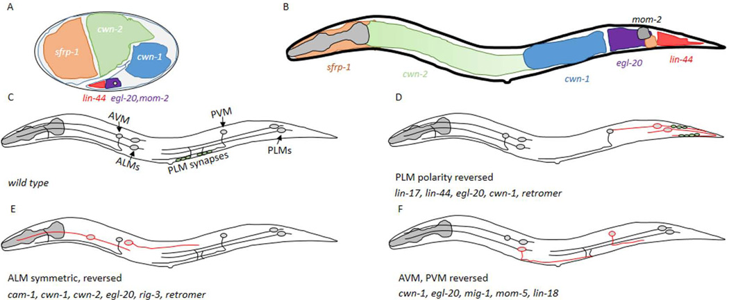
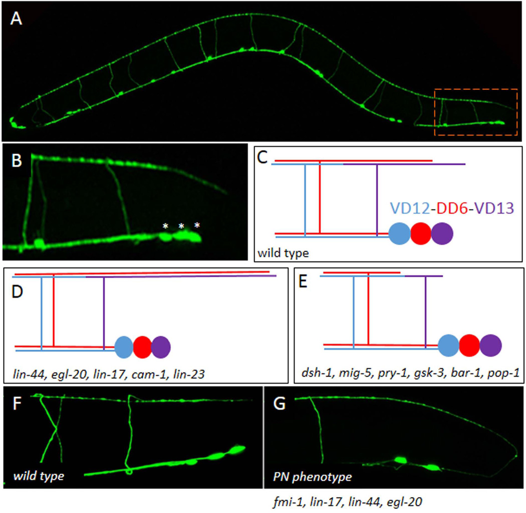
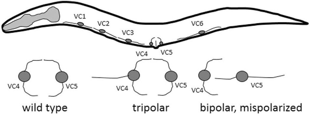
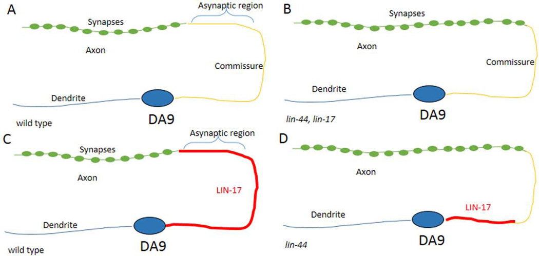
During the development of the nervous system, neurons encounter signals that inform their outgrowth and polarization. Understanding how these signals combinatorially function to pattern the nervous system is of considerable interest to developmental neurobiologists. The Wnt ligands and their receptors have been well characterized in polarizing cells during asymmetric cell division. The planar cell polarity (PCP) pathway is also critical for cell polarization in the plane of an epithelium. The core set of PCP genes include members of the conserved Wnt-signaling pathway, such as Frizzled and Disheveled, but also the cadherin-domain protein Flamingo. In Drosophila, the Fat and Dachsous cadherins also function in PCP, but in parallel to the core PCP components. C. elegans also have two Fat-like and one Dachsous-like cadherins, at least one of which, cdh-4, contributes to neural development. In C. elegans Wnt ligands and the conserved PCP genes have been shown to regulate a number of different events, including embryonic cell polarity, vulval morphogenesis, and cell migration. As is also observed in vertebrates, the Wnt and PCP genes appear to function to primarily provide information about the anterior to posterior axis of development. Here, we review the recent work describing how mutations in the Wnt and core PCP genes affect axon guidance and synaptogenesis in C. elegans.

Keywords: Disheveled; Flamingo; Frizzled; Wnt; planar cell polarity.


Figures

Figure 1
Figure 1. The mechanosensory neurons are dependent on Wnt signaling
A,B) A depiction of the areas that express the five different Wnt ligands during embryogenesis (A) and the L1 stage (B), based on in situ data from (Harterink et al., 2011). The grey circle in (A) indicates the approximate position of the PLM when axon outgrowth begins. Anterior is to the left and dorsal is up in all panels of this schematic. C) A cartoon of the six mechanosensory neurons present in C. elegans. The ALM neurons are in the anterior of the animal, the PLM neurons are posterior and the AVM and PVM cells are more centrally located. The position where PLM neurons make synapses is indicated by the green circles. D) In lin-17, lin-44, egl-20 mutants, or when the retromer complex was inactive, the PLM axons were polarized posteriorly. The axons were either symmetric or the posterior process was longer. The presynaptic protein, SNB-1 (green circles) was not aberrantly found posterior to the cell body. E) ALM neurons have migration and/or axon outgrowth defects in different Wnt signaling mutants. F) AVM and PVM make normal ventral projections, but have polarized growth errors in Wnt signaling mutants.
Figure 2
Figure 2. The GABAergic motoneurons use Wnts and FMI-1 to regulate A/P growth
A) The Punc-25∷gfp marker illuminates the four RME neurons in the head and the six DD and 13 VD motoneurons which are organized along the ventral midline. All 19 DD and VD neurons form commissures that project to the dorsal side of the animal resulting in a ladder-like appearance. B) In the tail the most posterior neurons VD12, DD6 and VD13 (asterisks) form a cluster. The dorsal branch of the VD13 axon projects to a point approximately even with the posterior edge of the cell cluster. C) A schematic of the VD12-DD6-VD13 cluster. D) Dorsal cord overgrowth due to mutations in the Wnt genes and receptors. E) Dorsal cord undergrowth was found in the downstream signaling proteins. F) In wild-type animals all the DD and VD neurons project anteriorly. G) An example of the PN defect present in the fmi-1 and Wnt signaling backgrounds.
Figure 3
Figure 3. The VC motoneurons have distinct morphologies
The six VC motoneurons are organized along the ventral midline, and normally have a bipolar appearance, with VC1, 2, 3 and 6 extending process anterior/posterior, while VC4 and 5 are rotated ~90 degrees to send process out left and right. Below, are examples of the phenotypes seen in the VC4 and VC5 neurons in the vang-1, prkl-1 and dsh-1 mutants, including extra neurites (tripolar) and a reversion to a more VC1-like bipolar appearance.
Figure 4
Figure 4. DA9 NMJs form in a LIN-17-dependent fashion
A) The DA9 neuron forms in the posterior of the animal, just anterior to the domain of LIN-44 expression (not shown). The cell body extends and anterior dendrite and a posterior process that forms an inverted C. Along the dorsal cord there is an asynaptic region, followed by a more anterior axonal process that forms NMJs. B) In the lin-17 or lin-44 mutants the asynaptic domain is shortened and synapses are formed closer to the commissure. C) In wild type the LIN-17 protein decorates the posterior process along the commissure and into the asynaptic domain, but not where synapses form. D) The LIN-17 accumulation to the commissure is lin-44 dependent.

References

    1. Ahmad FJ, Joshi HC, Centonze VE, Baas PW. Inhibition of microtubule nucleation at the neuronal centrosome compromises axon growth. Neuron. 1994;12:271–280. - PubMed
    1. Ambegaonkar AA, Pan G, Mani M, Feng Y, Irvine KD. Propagation of Dachsous-Fat planar cell polarity. Curr Biol. 2012;22:1302–1308. - PMC - PubMed
    1. Andersen EF, Halloran MC. Centrosome movements in vivo correlate with specific neurite formation downstream of LIM homeodomain transcription factor activity. Development. 2012;139:3590–3599. - PMC - PubMed
    1. Baas PW, Joshi HC. Gamma-tubulin distribution in the neuron: implications for the origins of neuritic microtubules. J Cell Biol. 1992;119:171–178. - PMC - PubMed
    1. Babu K, Hu Z, Chien SC, Garriga G, Kaplan JM. The immunoglobulin super family protein RIG-3 prevents synaptic potentiation and regulates Wnt signaling. Neuron. 2011;71:103–116. - PMC - PubMed

Publication types