. 2014 Jan;196(1):211-23.
doi: 10.1534/genetics.113.158485. Epub 2013 Nov 8.

Implicating SCF complexes in organogenesis in Caenorhabditis elegans

Affiliations

Implicating SCF complexes in organogenesis in Caenorhabditis elegans

Stanley R G Polley et al. Genetics. 2014 Jan.

Abstract

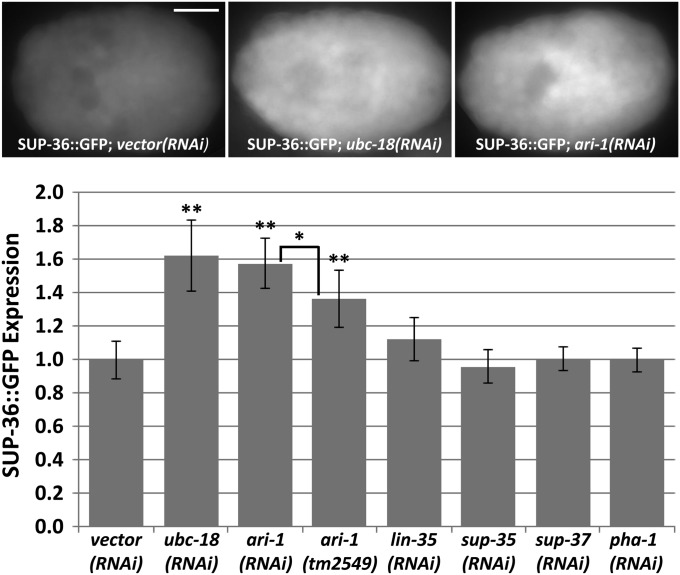
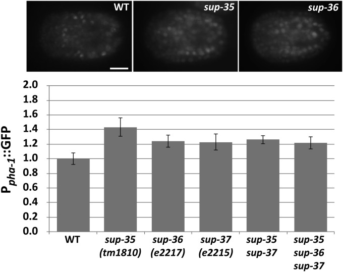
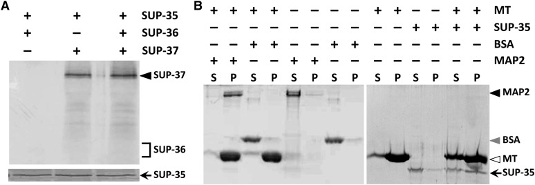
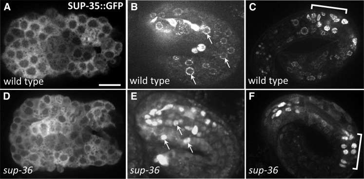
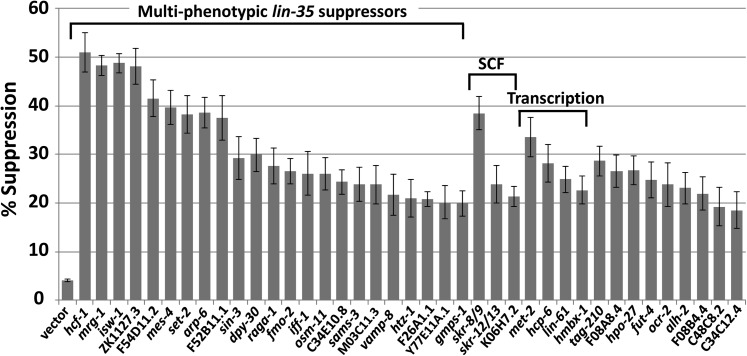
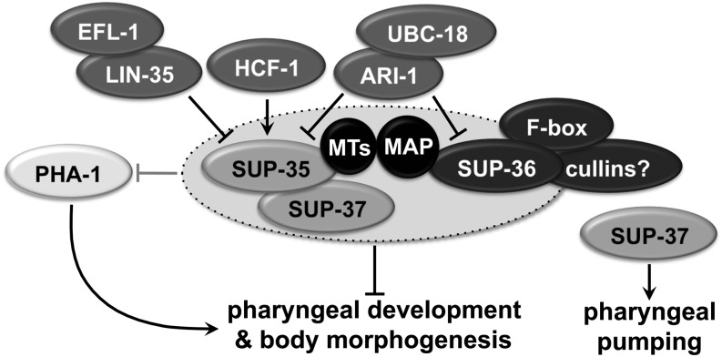
Development of the Caenorhabditis elegans foregut (pharynx) is regulated by a network of proteins that includes the Retinoblastoma protein (pRb) ortholog LIN-35; the ubiquitin pathway components UBC-18 and ARI-1; and PHA-1, a cytoplasmic protein. Loss of pha-1 activity impairs pharyngeal development and body morphogenesis, leading to embryonic arrest. We have used a genetic suppressor approach to dissect this complex pathway. The lethality of pha-1 mutants is suppressed by loss-of-function mutations in sup-35/ztf-21 and sup-37/ztf-12, which encode Zn-finger proteins, and by mutations in sup-36. Here we show that sup-36 encodes a divergent Skp1 family member that binds to several F-box proteins and the microtubule-associated protein PLT-1/τ. Like SUP-35, SUP-36 levels were negatively regulated by UBC-18-ARI-1. We also found that SUP-35 and SUP-37 physically associated and that SUP-35 could bind microtubules. Thus, SUP-35, SUP-36, and SUP-37 may function within a pathway or complex that includes cytoskeletal components. Additionally, SUP-36 may regulate the subcellular localization of SUP-35 during embryogenesis. We carried out a genome-wide RNAi screen to identify additional regulators of this network and identified 39 genes, most of which are associated with transcriptional regulation. Twenty-three of these genes acted via the LIN-35 pathway. In addition, several S-phase kinase-associated protein (Skp)1-Cullin-F-Box (SCF) components were identified, further implicating SCF complexes as part of the greater network controlling pharyngeal development.

Keywords: Caenorhabditis elegans; F-box; Skp1 pharynx; ari-1; lin-35; morphogenesis; pha-1; ptl-1; sup-35; sup-36; sup-37; ubc-18.


Figures

Figure 1
Figure 1
Pharyngeal phenotypes of pha-1 mutants and suppressed strains. (A–D) DIC micrographs of L1-stage larvae (A and C) and embryos (B and D) are shown. A and B show pha-1(ts) mutants grown at the nonpermissive temperatures of 22° and 25°, respectively. Note abnormal, compressed pharyngeal structures at both temperatures, whereas body morphogenesis defects are more severe at the higher temperature. C shows a suppressed pha-1(ts); sup-36(e2217) double mutant grown at 25° with a normal pharynx and body morphology. D shows a pha-1 embryo grown at the permissive temperature of 16° with overexpression of sup-36 by an extrachromosomal array (fdEx121). Open and solid arrowheads indicate anterior and posterior boundaries of the pharynx, respectively. Solid arrows delineate the pharyngeal lumen. Bar in A, 10 µm for A–D.
Figure 2
Figure 2
sup-36 genomic locus and SUP-36 protein sequence. (A) Schematic representation of the sup-36 genomic locus. Coding regions are indicated by regions with dark shading, and the 3′-UTR is shown as a region with light shading. Six sequenced sup-36 alleles are shown. e2217 and t1012 are deletion alleles (solid line) with endpoints that are not precisely defined (dashed lines). (B) Clustal alignment of SUP-36 and F15B10.3 from C. elegans, Skp1 from human, and two Skp1-like proteins from Encephalitozoon romaleae (microsporidia; AFN82639) and Ricinus communis (castor bean; XP_002513359). Solid blocks with open letters indicate identical amino acid residues; shaded blocks with solid letters indicate similar residues. Three missense mutations are indicated by asterisks. Arrowheads indicate boundaries of the conserved Skp1 dimerization domain.
Figure 3
Figure 3
SUP-36 Y2H interactions. (A) Results from a quantitative β-gal assay. Assays were carried out in triplicate, and error bars indicate 95% confidence intervals. Dashed line indicates the value of the highest negative control (sup-36/pDEST22). P-values were obtained using a Student’s t-test by comparing the indicated yeast strain to the control sup-36/pDEST22 (**P < 0.01, *P < 0.05). (B) Growth of the indicated yeast strains on His− (+25 mM 3-AT) and Ura− plates. C summarizes results for the four Y2H reporters. +++, strong reporter activation; +, weak interaction; and –, no interaction.
Figure 4
Figure 4
Embryonic expression pattern of SUP-36::GFP. (A–F) Confocal fluorescence images of full-length functional SUP-36::GFP in embryos. A and B show embryos prior to morphogenesis (∼100–350 min postfertilization), C and D show embryos at early stages of morphogenesis (∼350–400 min), E shows an embryo at the 1.5-fold stage (∼430 min), and F shows an ∼3-fold-stage embryo (>600 min). Arrows in A, B, and E indicate the positions of nuclei. Arrowheads in C indicate expression of SUP-36::GFP along the basal surface of primordial pharyngeal cells in the region abutting the basement membrane. Dotted outlined regions indicate the developing pharynx. Bar in A, 10 µm for A–F.
Figure 5
Figure 5
SUP-36::GFP expression regulation by upstream regulators. Expression levels of a full-length SUP-36::GFP reporter were assayed in embryos at the ∼300-cell stage under the indicated conditions. Top panels show representative fluorescence images of wild-type, ubc-18(RNAi), and ari-1(RNAi) embryos. Bottom panel shows quantification of embryos (n > 50 for each). Error bars indicate 95% confidence intervals (C.I.’s). Statistical analysis was carried out using Students t-test; **P < 0.001 relative to vector control, *P < 0.05. Bar, 10 µm.
Figure 6
Figure 6
pha-1 regulation by SUP-35, SUP-36, and SUP-37. Expression levels of a Ppha-1::GFP reporter were assayed in embryos at the ∼300-cell stage under the indicated conditions. Error bars indicate 95% C.I.’s. P < 0.05 based on Student’s t-test for all backgrounds relative to wild type.
Figure 7
Figure 7
SUP-35 interacts with both SUP-37 and microtubules. (A) Coprecipitation experiments using purified full-length His-tagged SUP-35 and full-length 35S-labeled SUP-36 and SUP-37 in vitro-translated proteins. The presence of correctly sized 35S-labeled SUP-36 and SUP-37 was confirmed in separate experiments (data not shown). SUP-36 and SUP-37 were detected by autoradiography, whereas SUP-35 was detected by Coomassie staining. (B) Microtubule (MT) pull-down binding assays that were carried out using full-length His-tagged SUP-35 along with positive (MAP2) and negative (BSA) controls as indicated. Note that the large majority of SUP-35 is contained within the supernatant (S) in the absence of microtubules but is enriched in the pellet fraction (P) in the presence of microtubules.
Figure 8
Figure 8
SUP-35::GFP localization is altered in sup-36 mutants. (A–F) Confocal fluorescence images of full-length functional SUP-35::GFP in embryos. A and D show embryos prior to morphogenesis (∼300–350 min postfertilization), B and E show twofold-stage embryos (∼450 min), and C and F show approximately threefold-stage embryos (>600 min). Both wild-type and sup-36(e2217) strains contained the same SUP-35::GFP extrachromosomal array (fdEx57). Arrows in B and E indicate the positions of nuclei. Brackets in C and F indicate the region containing posterior pharyngeal cells. Bar in A, 10 µm for A–F.
Figure 9
Figure 9
lin-35; ubc-18 suppressors identified by a whole-genome RNAi screen. Shown is percentage of suppression of larval arrest by 39 sequence-confirmed RNAi clones. Vector denotes a control RNAi. Suppressors have been organized into functional classes. The six new multiphenotypic lin-35 suppressor genes that were identified are arp-6, fmo-2, vamp-8, F26A1.1, Y77E11A.1, and gmps-1. Error bars indicate 95% C.I.’s. For all suppressor RNAi clones, P < 0.001 (relative to vector control) based on the exact test.
Figure 10
Figure 10
Model of pharyngeal regulation. Although binding between various binary components such as SUP-35–SUP-37 and SUP-35–microtubules was demonstrated by experimentation, it is unknown whether simultaneous interactions between multiple components also occur. For additional details, see text.

References

    1. Ahringer, J., 2005 Reverse genetics (April 6, 2006), WormBook, ed. The C. elegans Research Community, WormBook, /10.1895/wormbook.1.47.1, http://www.wormbook.org.
    1. Andersen E. C., Horvitz H. R., 2007. Two C. elegans histone methyltransferases repress lin-3 EGF transcription to inhibit vulval development. Development 134: 2991–2999. - PubMed
    1. Andersen E. C., Saffer A. M., Horvitz H. R., 2008. Multiple levels of redundant processes inhibit Caenorhabditis elegans vulval cell fates. Genetics 179: 2001–2012. - PMC - PubMed
    1. Anderson D. D., Eom J. Y., Stover P. J., 2012. Competition between sumoylation and ubiquitination of serine hydroxymethyltransferase 1 determines its nuclear localization and its accumulation in the nucleus. J. Biol. Chem. 287: 4790–4799. - PMC - PubMed
    1. Bessler J. B., Andersen E. C., Villeneuve A. M., 2010. Differential localization and independent acquisition of the H3K9me2 and H3K9me3 chromatin modifications in the Caenorhabditis elegans adult germ line. PLoS Genet. 6: e1000830. - PMC - PubMed

Publication types

MeSH terms