. 2014 Jan;196(1):197-210.
doi: 10.1534/genetics.113.156281. Epub 2013 Nov 8.

Dual phosphorylation of cdk1 coordinates cell proliferation with key developmental processes in Drosophila

Affiliations

Dual phosphorylation of cdk1 coordinates cell proliferation with key developmental processes in Drosophila

Joseph O Ayeni et al. Genetics. 2014 Jan.

Abstract

Eukaryotic organisms use conserved checkpoint mechanisms that regulate Cdk1 by inhibitory phosphorylation to prevent mitosis from interfering with DNA replication or repair. In metazoans, this checkpoint mechanism is also used for coordinating mitosis with dynamic developmental processes. Inhibitory phosphorylation of Cdk1 is catalyzed by Wee1 kinases that phosphorylate tyrosine 15 (Y15) and dual-specificity Myt1 kinases found only in metazoans that phosphorylate Y15 and the adjacent threonine (T14) residue. Despite partially redundant roles in Cdk1 inhibitory phosphorylation, Wee1 and Myt1 serve specialized developmental functions that are not well understood. Here, we expressed wild-type and phospho-acceptor mutant Cdk1 proteins to investigate how biochemical differences in Cdk1 inhibitory phosphorylation influence Drosophila imaginal development. Phosphorylation of Cdk1 on Y15 appeared to be crucial for developmental and DNA damage-induced G2-phase checkpoint arrest, consistent with other evidence that Myt1 is the major Y15-directed Cdk1 inhibitory kinase at this stage of development. Expression of non-inhibitable Cdk1 also caused chromosome defects in larval neuroblasts that were not observed with Cdk1(Y15F) mutant proteins that were phosphorylated on T14, implicating Myt1 in a novel mechanism promoting genome stability. Collectively, these results suggest that dual inhibitory phosphorylation of Cdk1 by Myt1 serves at least two functions during development. Phosphorylation of Y15 is essential for the premitotic checkpoint mechanism, whereas T14 phosphorylation facilitates accumulation of dually inhibited Cdk1-Cyclin B complexes that can be rapidly activated once checkpoint-arrested G2-phase cells are ready for mitosis.

Keywords: Cdk1; cell-cycle checkpoint; imaginal discs; mitosis; neuroblast.


Figures

Figure 1
Figure 1
Biochemical analysis of transgenic Cdk1 fusion proteins expressed in wing discs with en-Gal. In A and C, each sample was extracted from 10 late third instar wing discs. (A) Western blot of wing disc extracts probed with PSTAIRE antibodies (conserved Cdk1 epitope) detected both endogenous (34 kDa) and transgenic fusion (61 kDa) proteins, except in the nontransgenic yw control. Adjusting for exposure times, we estimated that levels of the transgenic Cdk1 proteins were approximately fourfold lower than endogenous Cdk1 in this experiment. (B) Western blot of transgenic Cdk1 that was immunoprecipitated from wing disc lysates (30–35 discs per sample) using rabbit anti-GFP. The blot was sequentially probed with mouse anti-GFP, Drosophila Cyclin B, and Cyclin A. Note that Cyclin A runs as a doublet (Lehner and O’Farrell 1990). (C) Western blot of wing-disc extracts with the top section (proteins above 48 kDa) probed sequentially with phospho-specific antibodies against the pT14-Cdk1, GFP, pY15-Cdk1, and pT161-Cdk1 phospho-isoforms, stripping between each reprobing. The bottom part of the blot containing 34-kDa endogenous Cdk1 was probed with PSTAIRE antibodies as a loading control. (D) Compilation of data from three independent experiments for each genotype that measured histone H1 kinase activity of Cdk1 fusion proteins immunoprecipitated from wing-disc extracts with anti-GFP antibodies (180 discs for each sample). We determined kinase activity for each Cdk1 variant by normalizing the H1 kinase activity per unit of total Cdk1 protein, estimated by probing Western blots of aliquots of the immunoprecipitates with anti-PSTAIRE for each experiment. Relative percentage kinase activity was plotted with respect to Cdk1(WT) activity, which was set as 100%. Error bars show standard deviation calculated from data from three experimental replicates for each genotype.
Figure 2
Figure 2
Temperature-sensitive cdc2 (cdk1) mutants are rescued by ubiquitous expression of Cdk1WT and Cdk1(T14A), but not by Cdk1(Y15F) or Cdk1(T14AY15F). (A and A′) Dorsal and side views of wild-type control flies, respectively. (B and B′) Flies ubiquitously expressing the Cdk1WT-VFP transgene with tubulin-Gal4 in temperature-sensitive cdc2 mutant background (cdc2B47/cdc2E1-E24;UAS-Cdk1WT-VFP/tubulin-Gal4). (C and C′) Adult flies ubiquitously expressing the Cdk1(T14A)-VFP transgene with tubulin-Gal4 (cdc2B47/cdc2E1-E24;UAS-Cdk1(T14A)-VFP/Gal4). (D) Summary of progeny data tabulated in Figure S1E, showing the percentage of rescued flies relative to the expected maximum percentage rescue.
Figure 3
Figure 3
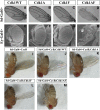
Phenotypic effects of expression of Cdk1 transgenes during adult wing and eye development are enhanced by co-expression of Cyclin B using sd-Gal4 and ey-Gal4, respectively, with ∼500 adults examined for each genotype. (A–C) Progeny expressing Cdk1(WT), Cdk1(T14A), or Cdk1(Y15F) in an otherwise wild-type background that have no defects in adult wing morphology. (D) Cdk1(T14A,Y15F) expression caused wing-margin defects. (E–G) Scanning electron micrographs of adult eyes from progeny expressing Cdk1(WT), Cdk1(T14A), or Cdk1(Y15F) that did not affect adult eye development. (H) Cdk1(T14A,Y15F) expression severely affected adult eye and head structures and caused pharate adult lethality (n = 200). (I–K) Expression of Cyclin B alone, co-expression of Cyclin B with Cdk1(WT), or co-expression of Cyclin B with Cdk1(T14A) had no detectable effect on adult wings. (L) Co-expression of Cdk1(Y15F) with Cyclin B caused extensive loss of adult wing margin. (M) Co-expression of Cdk1(T14A,Y15F) with Cyclin B resulted in complete loss of the adult wing.
Figure 4
Figure 4
Expression of Cdk1(Y15F) and Cdk1(T14A,Y15F) caused increased apoptosis and mitotic index in late third instar larval wing discs. VFP-tagged Cdk1 constructs (green label in E–H and M–P) were expressed with en-Gal4 in the posterior compartment, and the discs were labeled with antibodies against activated Caspase-3 (red in E–H) as an apoptosis marker or antibodies against phosphorylated (S10) histone H3 (PH3; red in M, N, O, and P) as a mitotic marker. The VFP-negative anterior compartment serves as an internal control. (A, B, E, and F) Few apoptotic cells were observed in discs expressing Cdk1(WT) or Cdk1(T14A) (n =10, each genotype). (C, D, G, and H) Expression of Cdk1(Y15F) or Cdk1(T14A,Y15F) resulted in elevated numbers of apoptotic cells in the posterior region of wing discs (n = 10). (I, J, M, and N) No notable differences in PH3-labeled cells from expression of Cdk1(WT) or Cdk1(T14A) in wing discs (n = 15, each genotype). (K, L, O, and P) More PH3-labeled cells were observed in posterior regions of Cdk1(Y15F)- and Cdk1(T14A,Y15F)-expressing discs (n = 9, each genotype). Wing discs expressing Cdk1(T14A,Y15F) were always smaller and morphologically abnormal relative to the other genotypes, as shown. Bar in A, 50 µm.
Figure 5
Figure 5
Expression of Cdk1 transgenes during wing development with sd-Gal4 differentially affects wing-margin pattern and developmental regulation of G2-phase arrest. At least 10 labeled discs were examined for each genotype. (A–C) Wing discs from third instar larvae labeled with anti-Cut antibodies (red). Cut expression at the presumptive wing margin was unaffected by expression of Cdk1(WT), Cdk1(T14A), or Cdk1(Y15F), respectively. (D) Expression of Cdk1(T14A,Y15F) disrupted Cut expression. (A′–D′) VFP-tagged transgenes were expressed throughout the wing pouch of each disc. (E–H) Wing discs from third instar larvae expressing transgenes driven by neurp72-Gal4 in SOP cells (a subset of G2-phase-arrested ZNC cells) that were labeled with antibodies against phosphorylated (S10) histone H3 (PH3; red). (E and F) SOP cells expressing Cdk1(WT)-VFP and Cdk1(T14A) were PH3-negative. (G and H) SOP cells expressing Cdk1(Y15F)-VFP and Cdk1(T14A,Y15F) were a mixture of smaller mitotic (PH3-positive) and nonmitotic cells. (E′–H′) VFP-tagged transgenes were expressed in SOP cells.
Figure 6
Figure 6
Expression of Cdk1(Y15F)-VFP and Cdk1(T14A,Y15F) in wing discs causes dominant bypass of DNA-damage-induced G2/M checkpoint responses. VFP-tagged Cdk1 transgenes (green) were expressed with en-Gal4 in the posterior compartment of each wing disc. Wing discs were dissected from late third instar larvae 60 min after exposure to 40 Gy of ionizing radiation and labeled with antibodies against phosphorylated (S10) histone H3 (PH3; white in A–D, red in A′–D′) to mark mitotic cells. At least 10 labeled discs were examined for each genotype. (A, A′, B, and B′) No PH3-positive cells were in either compartment of wing discs expressing Cdk1(WT) or Cdk1(T14A), indicating a functional premitotic checkpoint response. (C, C′, D, and D′) Wing discs expressing Cdk1(Y15F) or Cdk1(T14A,Y15F), where PH3 labeling in the posterior compartment indicated a G2/M checkpoint defect. Bar in A, 50 µm.
Figure 7
Figure 7
Expression of Cdk1(Y15F)-VFP and Cdk1(T14A,Y15F) with prospero-Gal4 differentially affects mitotic index measurements in type 1 larval neuroblasts; however, only Cdk1(T14A,Y15F) produced gross chromosome defects. Metaphase karyotypes of colchicine-treated brain squashes were labeled with Hoechst 33258 to identify mitotic chromosomes. At least 800 interpretable karyotypes were examined for each genotype. (A) Bar chart showing mitotic index associated with each Cdk1 transgene. (B and C) Neuroblasts expressing Cdk1(WT) or Cdk1(T14A) have chromosomes arrested in metaphase with cohered sister chromatids. (D) Approximately 90% of Cdk1(Y15F)-expressing mitotic neuroblasts also arrest in metaphase with cohered sister chromatids; however, defects in sister-chromatid cohesion were evident in 9.5% of the karyotypes (E, n = 900). Approximately 45% of Cdk1(T14A,Y15F)-expressing neuroblasts exhibited some form of chromosomal aberration (n = 850), including polyploidy (G); thin, poorly condensed chromosomes (H); or chromosome breaks (arrow).
Figure 8
Figure 8
Live imaging of mitosis in larval neuroblasts driven by prospero-Gal4 of Cdk1-VFP (green) and tubulin-RFP (red) shows that only Cdk1(T14A,Y15F) expression causes mitotic timing defects. Each set of panels represent still images taken at four time points from each of the movies in File S1, File S2, File S3, and File S4. (A–D) (Top) Merged images of the respective Cdk1-VFP (green) and tubulin-RFP (red) fluorescent reporters. (Bottom) Only tubulin-RFP. In A–D, the zero time point (first panels) marks the appearance of centrosomes at opposite poles (t = 0); the second panels show midpoint of bipolar spindle formation; and the third and fourth panels represent the beginning and end of cytokinesis, respectively. (A) Neuroblasts expressing Cdk1WT-VFP, where average time of mitosis was 12.56 ± 1.2 min (n = 5) (File S1). (B) Neuroblasts expressing Cdk1(T14A), where average time of mitosis was 13.52 ± 1.26 min (n = 5) (File S2). (C) Neuroblasts expressing Cdk1(Y15F), where average time of mitosis was 13.0 ± 0.7 min (n = 5) (File S3). (D) Neuroblasts expressing Cdk1(T14A,Y15F), where average time of mitosis was 28.54 ± 2.25 min, (n = 3) (File S4). (E) Quantification of the mitotic timing data for neuroblasts expressing Cdk1WT-VFP (n = 5), Cdk1(T14A)-VFP (n = 5), Cdk1(Y15F)-VFP (n = 5), and Cdk1(T14A,Y15F)-VFP (n = 3). The bar graph depicts the mean duration of mitosis ± SD for each genotype.

References

    1. Basto R., Gomes R., Karess R. E., 2000. Rough deal and Zw10 are required for the metaphase checkpoint in Drosophila. Nat. Cell Biol. 2: 939–943. - PubMed
    1. Blasina A., Paegle E. S., McGowan C. H., 1997. The role of inhibitory phosphorylation of CDC2 following DNA replication block and radiation-induced damage in human cells. Mol. Biol. Cell 8: 1013–1023. - PMC - PubMed
    1. Booher R. N., P. S. Holman, and A. Fattaey, 1997. Human Myt1 is a cell cycle-regulated kinase that inhibits Cdc2 but not Cdk2 activity. J. Biol. Chem. 272: 22300–22306. - PubMed
    1. Brand A. H., and N. Perrimon, 1993. Targeted gene expression as a means of altering cell fates and generating dominant phenotypes Development 118: 401–415. - PubMed
    1. Brodsky M. H., W. Nordstrom, G. Tsang, E. Kwan, G. M. Rubin et al, 2000. Drosophila p53 binds a damage response element at the reaper locus. Cell 101: 103–113. - PubMed

Publication types