

High-resolution diffusion-weighted imaging for the separation of benign from malignant BI-RADS 4/5 lesions found on breast MRI at 3T
- PMID: 24214467
- PMCID: PMC4014534
- DOI: 10.1002/jmri.24416
High-resolution diffusion-weighted imaging for the separation of benign from malignant BI-RADS 4/5 lesions found on breast MRI at 3T
Abstract
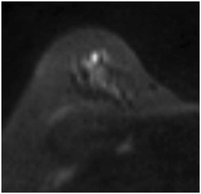
Purpose: To determine whether readout-segmented echo-planar diffusion imaging (RESOLVE) improves separation of malignant versus benign lesions compared to standard single-shot echo-planar imaging (ss-EPI) on BI-RADS 4/5 lesions detected on breast magnetic resonance imaging (MRI).
Materials and methods: Consecutive 3T breast MRI studies with BI-RADS 4/5 designation and subsequent biopsy or benign mastectomy were retrospectively identified. Freehand regions of interest (ROIs) were drawn on lesions and also on normal background fibroglandular tissue for comparison. Lesion-to-background contrast was evaluated by normalizing signal intensity of the lesion ROI by the normal background tissue ROI at b = 800. Statistical analysis used the Mann-Whitney/Wilcoxon rank-sum test for unpaired and Wilcoxon signed-rank for paired comparisons.
Results: Of 38 lesions in 32 patients, 10 were malignant. Lesion-to-background contrast was higher on RESOLVE than ss-EPI (1.80 ± 0.71 vs. 1.62 ± 0.63, P = 0.03). Mean apparent diffusion coefficient (ADC) was the same or lower on RESOLVE than ss-EPI, and this effect was largest in malignant lesions (RESOLVE 0.90 ± 0.13; ss-EPI 1.00 ± 0.13; median difference -0.10 (95% confidence interval [CI]: -0.17, -0.02) × 10(-3) mm(2) /sec; P = 0.014). By either diffusion method, there was a statistically significant difference between benign and malignant mean ADC (P < 0.001).
Conclusion: Increased lesion-to-background contrast and improved separation of benign from malignant lesions by RESOLVE compared to standard diffusion suggests that RESOLVE may show promise as an adjunct to clinical breast MRI.
Keywords: DWI; benign; breast MRI; diffusion; malignant; screening.
© 2013 Wiley Periodicals, Inc.
Figures


References
-
- Kuhl CK, Schrading S, Bieling HB, Wardelmann E, Leutner CC, Koenig R, et al. MRI for diagnosis of pure ductal carcinoma in situ: a prospective observational study. Lancet. 2007 Aug 11;370(9586):485–92. - PubMed
-
- Medeiros LR, Duarte CS, Rosa DD, Edelweiss MI, Edelweiss M, Silva FR, et al. Accuracy of magnetic resonance in suspicious breast lesions: a systematic quantitative review and meta-analysis. Breast Cancer Res Treat. 2011 Apr;126(2):273–85. - PubMed
-
- DeMartini W, Lehman C, Partridge S. Breast MRI for cancer detection and characterization: a review of evidence-based clinical applications. Acad Radiol. 2008 Apr;15(4):408–16. - PubMed
-
- Kul S, Cansu A, Alhan E, Dinc H, Gunes G, Reis A. Contribution of diffusion-weighted imaging to dynamic contrast-enhanced MRI in the characterization of breast tumors. AJR American journal of roentgenology. 2011 Jan;196(1):210–7. - PubMed
-
- Partridge SC, Mullins CD, Kurland BF, Allain MD, DeMartini WB, Eby PR, et al. Apparent diffusion coefficient values for discriminating benign and malignant breast MRI lesions: effects of lesion type and size. AJR American journal of roentgenology. 2010 Jun;194(6):1664–73. - PubMed
Publication types
MeSH terms
Substances
Grants and funding
LinkOut - more resources
Full Text Sources
Other Literature Sources
Medical
