. 2014 Jan;164(1):201-15.
doi: 10.1104/pp.113.228338. Epub 2013 Nov 8.

RBF1, a plant homolog of the bacterial ribosome-binding factor RbfA, acts in processing of the chloroplast 16S ribosomal RNA

Affiliations

RBF1, a plant homolog of the bacterial ribosome-binding factor RbfA, acts in processing of the chloroplast 16S ribosomal RNA

Rikard Fristedt et al. Plant Physiol. 2014 Jan.

Erratum in

  • CORRECTIONS.
    [No authors listed] [No authors listed] Plant Physiol. 2015 Oct;169(2):1419. doi: 10.1104/pp.15.01381. Plant Physiol. 2015. PMID: 26417055 Free PMC article. No abstract available.

Abstract

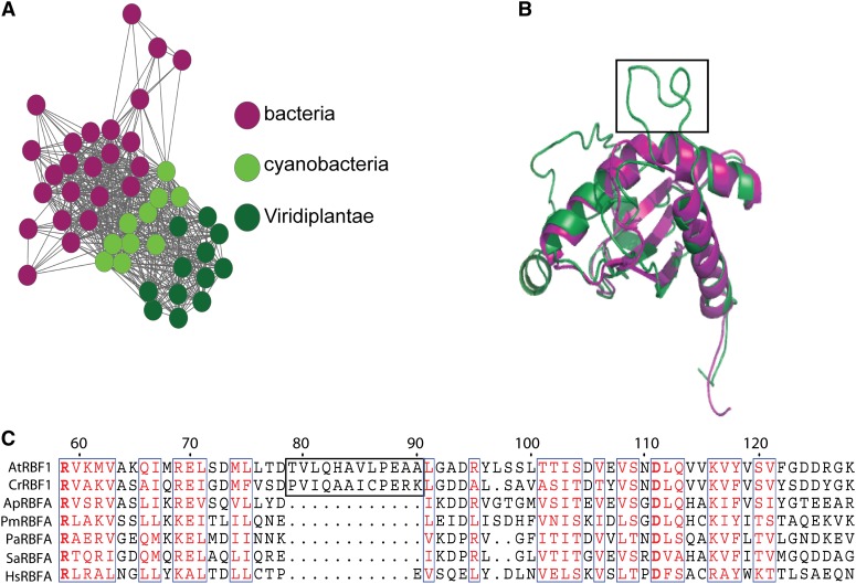
Plastids (chloroplasts) possess 70S ribosomes that are very similar in structure and function to the ribosomes of their bacterial ancestors. While most components of the bacterial ribosome (ribosomal RNAs [rRNAs] and ribosomal proteins) are well conserved in the plastid ribosome, little is known about the factors mediating the biogenesis of plastid ribosomes. Here, we have investigated a putative homolog of the bacterial RbfA (for ribosome-binding factor A) protein that was identified as a cold-shock protein and an auxiliary factor acting in the 5' maturation of the 16S rRNA. The unicellular green alga Chlamydomonas reinhardtii and the vascular plant Arabidopsis (Arabidopsis thaliana) both encode a single RbfA-like protein in their nuclear genomes. By generating specific antibodies against this protein, we show that the plant RbfA-like protein functions exclusively in the plastid, where it is associated with thylakoid membranes. Analysis of mutants for the corresponding gene (termed RBF1) reveals that the gene function is essential for photoautotrophic growth. Weak mutant alleles display reduced levels of plastid ribosomes, a specific depletion in 30S ribosomal subunits, and reduced activity of plastid protein biosynthesis. Our data suggest that, while the function in ribosome maturation and 16S rRNA 5' end processing is conserved, the RBF1 protein has assumed an additional role in 3' end processing. Together with the apparent absence of a homologous protein from plant mitochondria, our findings illustrate that the assembly process of the 70S ribosome is not strictly conserved and has undergone some modifications during organelle evolution.


Figures

Figure 1.
Figure 1.
RBF1 is related to but distinct from bacterial RbfA proteins. A, Protein similarity network of RbfA domain proteins. RbfA domain protein sequences from bacteria, cyanobacteria, green algae, and embryophyte plants were compared in an all-versus-all BLAST with a cutoff e-value of 10−18 and visualized with Cytoscape (http://www.cytoscape.org/). Each circle represents one protein. Lines connect proteins that have e-values below the cutoff. B, Comparison of RbfA structures. The predicted structure of the RBFA protein from Arabidopsis (green) is superimposed on the crystal structure of RbfA from the thermophile bacterium Thermotoga maritima (Protein Data Bank no. 2KZF; pink). I-TASSER was used to generate the predicted structure (Roy et al., 2010). The boxed region represents the Viridiplantae-specific insertion and corresponds to the boxed sequence in C. C, Amino acid sequence alignment of RbfA-like proteins from Arabidopsis (At), C. reinhardtii (Cr), Arthrospira platensis (Ap), Prochlorococcus marinus strain MIT 9215 (Pm), Pseudomonas aeruginosa MPAO1/P2 (Pa), Staphylococcus aureus subspecies aureus N315 (Sa), and Homo sapiens (Hs), where RBFA is predicted to be a mitochondrial protein (http://www.ncbi.nlm.nih.gov/gene/79863). The region boxed in black (amino acids 120–132) represents the plant-specific insertion (see B) that is absent from all bacterial RbfA proteins. Highly conserved amino acids are boxed in blue and indicated in red font, and absolutely conserved ones are in boldface red font. Only the conserved domain of RbfA is shown in this alignment. For a complete sequence alignment, see Supplemental Figure S1.
Figure 2.
Figure 2.
RBF1 is expressed early in plant development and is loosely associated with the thylakoid membrane. A, Domain organization and exon-intron structure of the RBF1 gene. The RBF1 genomic locus contains three exons (dark gray boxes) and two introns (black lines). Light gray boxes represent the UTRs (5′ and 3′), and the white box denotes the putative transit peptide (TP) for protein import into plastids. The region used to generate RBF1-specific antisera is indicated. B, RBF1 protein accumulation during plant development. Thylakoid membrane samples equivalent to 1 μg of chlorophyll from 1- to 6-week-old Arabidopsis plants were loaded as indicated and analyzed by immunoblotting with anti-RBF1, anti-Lhcb1.1, and anti-Lhcb1.2 antisera. A representative blot stained with Ponceau S is shown at the bottom. C, Analysis of the subcellular localization of the RBF1 protein in Arabidopsis. Total plant extract (Ex) and nuclear, mitochondrial, chloroplast, thylakoid, and chloroplast stromal fractions (Nucl, Mito, Cp, T, and S) were purified, and equivalent amounts of protein (10 μg per sample) were analyzed by immunoblotting using antibodies against RBF1 and the marker proteins D1 (a reaction center protein of PSII), cytochrome f (Cyt f; a subunit of the cytochrome b6f complex in the thylakoid membrane), the mitochondrial outer membrane protein TOM40, the nuclear protein CRYPTOCHROME2 (CRY2), and the soluble chloroplast protein RbcL (for large subunit of Rubisco). D, Weak membrane association of chloroplast RBF1. Isolated thylakoid membranes were washed with 0.4 m NaCl, and the thylakoid membranes (T) and the supernatant (S) were probed by immunoblotting with antibodies against RBF1 and the PSII reaction center protein D1.
Figure 3.
Figure 3.
Isolation of Arabidopsis rbf1 mutants, and characterization of their phenotypes. A, T-DNA insertion sites in tagged Arabidopsis rbf1 mutants. T-DNA insertion sites (black triangles) are shown in relation to the RBF1 gene structure. The three rbf1 alleles analyzed in this study are denoted as rbf1-1, rbf1-2, and rbf1-3. The RBF1 coding region is indicated by the translational start (ATG) and stop (TAA) codons. The exon-intron structure of the RBF1 genomic locus is represented as in Figure 2. B, Immunoblot detection of RBF1 in leaf extracts of wild-type (WT) and rbf1 mutant plants using anti-RBF1 antibodies. Leaf total protein extracts from 3-week-old wild-type and rbf1 seedlings (for growth conditions, see “Materials and Methods”; 10 μg of protein loaded) were separated by SDS-PAGE and probed with anti-RBF1 antibodies. A representative blot stained with Ponceau S is shown at the bottom. C, Phenotypes of rbf1 mutants. The top row shows, from left to right, a wild-type plant, an rbf1-1 mutant plant (exhibiting pale young leaves), and a complemented rbf1-1 mutant plant (rbf1-C). The bottom row shows, from left to right, a wild-type plant, an rbf1-2 mutant plant, and an rbf1-3 mutant plant grown on Suc-containing synthetic medium. Plants were grown under long-day conditions (16 h of light, 8 h of dark) at a photon flux density of 120 μmol m−2 s−1. Bars = 1 cm.
Figure 4.
Figure 4.
Comparison of chloroplast ultrastructure in wild-type (WT) and rbf1-1 mutant plants. Young leaves from wild-type and rbf1-1 mutant plants were analyzed by electron microscopy. A, Chloroplast ultrastructure. Note the strongly reduced grana stacking in the rbf1-1 mutant. B, Enlargement of the region boxed in A showing the structure of thylakoid membranes in the wild type and the mutant.
Figure 5.
Figure 5.
Thylakoid protein accumulation and photosynthesis in the rbf1-1 mutant. A, Immunoblot analysis of thylakoid proteins diagnostic for PSII, PSI, and ATP synthase. For quantitative comparison, a dilution series for the wild type (WT; 25%, 50%, and 100%) was loaded. Protein extracts were separated by SDS-PAGE and probed with specific antibodies directed against D1 (PSII reaction center subunit), Lhcb1 (PSII antenna protein), PsaA (PSI reaction center subunit), Lhca1 (PSI antenna protein), CF1αβ (ATP synthase subunits), and the large subunit of Rubisco (RbcL), a soluble protein in the stroma. A representative gel stained with Coomassie blue is shown below the blots to confirm even loading of the gel. B, Blue-native gel electrophoretic analysis of thylakoidal protein complexes from young (YL) and mature (ML) leaves. C, False-color display of Fv/Fm of rbf1-1 and wild-type plants. The color scale representing Fv/Fm is given at the right.
Figure 6.
Figure 6.
Accumulation of rRNAs as a proxy for the corresponding ribosomal subunits of the cytosolic and plastid ribosomes. The ratios of plastid 16S rRNA (component of the 30S subunit) to plastid 23S rRNA (component of the 50S subunit), plastid rRNAs to the cytosolic 18S rRNA, and cytosolic 18S rRNA to cytosolic 25S rRNA are shown for the wild type (WT) and the rbf1-1 and rbf1-2 mutants. “23SH1” is the largest “hidden break” product of the 23S rRNA (Delp and Kössel, 1991; Tiller et al., 2012), and “23S” is the 2.8-kb precursor. Significant differences (P < 0.05) are marked with asterisks. Data are from three technical replicates for each plant line, and error bars indicate sd. A, rbf1-1 young leaves. B, rbf1-1 mature (fully expanded) leaves. C, rbf1-2 mature (fully expanded) leaves.
Figure 7.
Figure 7.
Analysis of rRNA processing in rbf1 mutant plants. A, Physical map and transcript pattern of the plastid rRNA operon. The 7.3-kb primary transcripts, the various processing intermediates, and the mature forms of the 16S and 23S rRNAs are shown. Note that the 23S rRNA is cleaved into three pieces, a phenomenon known as hidden break processing (Delp and Kössel, 1991). The positions of the hybridization probes for both genes are also indicated. B, Analysis of 16S rRNA accumulation in wild-type plants (WT) and the rbf1-1 and rbf1-2 mutants. Young and mature leaves from soil-grown plants were analyzed for the rbf1-1 mutant (which grows autotrophically; Fig. 3), whereas plant material grown on Suc-containing synthetic medium (in vitro) was analyzed for the rbf1-2 mutant (because of its seedling-lethal phenotype; Fig. 3; Supplemental Fig. S3). As a loading control, the ethidium bromide-stained gel region containing the cytosolic 25S rRNA is shown. C, Analysis of 23S rRNA accumulation by RNA gel-blot hybridization. The same samples used in B were analyzed.
Figure 8.
Figure 8.
Detection of 5′ unprocessed and 3′ unprocessed precursors of the 16S rRNA. RNA blots were hybridized to probes specifically recognizing 16S precursors with 5′ extensions (5′ precursor; top) or 3′ extensions (3′ precursor; bottom; Supplemental Table S1). The rbf1-2 mutant and the corresponding wild type (WT) were grown on synthetic medium (due to the severe phenotype of the rbf1-2 mutant; compare with Supplemental Fig. S3), whereas the rbf1-1 mutant and its wild-type control were grown in soil. As a loading control, the ethidium bromide-stained gel region containing the cytosolic 25S rRNA is shown.
Figure 9.
Figure 9.
Distribution of plastid rRNAs and rRNA precursors between free ribosomes and polysomes in the rbf1-1 mutant and the wild type (WT). Free ribosomes and polysomes were separated using Suc gradient centrifugation, followed by RNA isolation and gel-blot analysis with probes specific for plastid rRNAs and their precursors. The wedges at the bottom indicate the increasing Suc density in the gradients. The light gradient fractions (left lanes) are enriched in free ribosomes, whereas the heavy fractions (right lanes) are enriched in polysomes. The methylene blue-stained cytosolic 18S rRNA is shown as a loading control below each blot. A, Hybridization to a 16S rRNA-specific probe (compare with Fig. 7A). Note the stronger presence of precursor molecules in the rbf1-1 mutant (best visible in fractions 1–3) and the underrepresentation of the 16S rRNA in the heaviest fraction (fraction 6). B, Hybridization to a 16S rRNA precursor-specific probe. The probe hybridizes downstream of the mature 3′ end (nucleotide positions +39 to +116). C, Hybridization to a 23S rRNA-specific probe (compare with Fig. 7A). D, Hybridization to a 23S rRNA precursor-specific probe. The probe binds upstream of the mature 5′ end (nucleotide positions −100 to −26). E, Identification of polysome-containing gradient fractions by centrifugation in the presence of the polysome-dissociating agent puromycin. The blot was hybridized to the 16S rRNA-specific probe.
Figure 10.
Figure 10.
Polysome loading of selected plastid mRNAs in the wild type (WT) and the rbf1-1 mutant. Blot hybridization analyses of RNAs extracted from fractionated polysome gradients are shown. The methylene blue-stained cytosolic 18S rRNA is shown as a loading control below each blot. The wedges at the bottom indicate the increasing Suc density in the gradient. A, Hybridization to an rbcL-specific probe. Note that the rbcL mRNA peaks in fraction 5 in the wild type but peaks already in fraction 4 in the mutant. B, Hybridization to a psbA-specific probe. C, Hybridization to a psbD-specific probe. Note that the psbD mRNA peaks in fraction 5 in the wild type but in fractions 4 and 5 in the mutant. D, Identification of polysome-containing gradient fractions by centrifugation in the presence of the polysome-dissociating agent puromycin. The blot was hybridized to the rbcL-specific probe.

References

    1. Albrecht V, Ingenfeld A, Apel K. (2006) Characterization of the snowy cotyledon 1 mutant of Arabidopsis thaliana: the impact of chloroplast elongation factor G on chloroplast development and plant vitality. Plant Mol Biol 60: 507–518 - PubMed
    1. Alkatib S, Scharff LB, Rogalski M, Fleischmann TT, Matthes A, Seeger S, Schöttler MA, Ruf S, Bock R. (2012) The contributions of wobbling and superwobbling to the reading of the genetic code. PLoS Genet 8: e1003076. - PMC - PubMed
    1. Arabidopsis Genome Initiative (2000) Analysis of the genome sequence of the flowering plant Arabidopsis thaliana. Nature 408: 796–815 - PubMed
    1. Archibald JM. (2009) The puzzle of plastid evolution. Curr Biol 19: R81–R88 - PubMed
    1. Asakura Y, Galarneau E, Watkins KP, Barkan A, van Wijk KJ. (2012) Chloroplast RH3 DEAD box RNA helicases in maize and Arabidopsis function in splicing of specific group II introns and affect chloroplast ribosome biogenesis. Plant Physiol 159: 961–974 - PMC - PubMed

Publication types

MeSH terms