. 2014 Jan;164(1):105-18.
doi: 10.1104/pp.113.230185. Epub 2013 Nov 8.

Leaf oil body functions as a subcellular factory for the production of a phytoalexin in Arabidopsis

Affiliations

Leaf oil body functions as a subcellular factory for the production of a phytoalexin in Arabidopsis

Takashi L Shimada et al. Plant Physiol. 2014 Jan.

Abstract

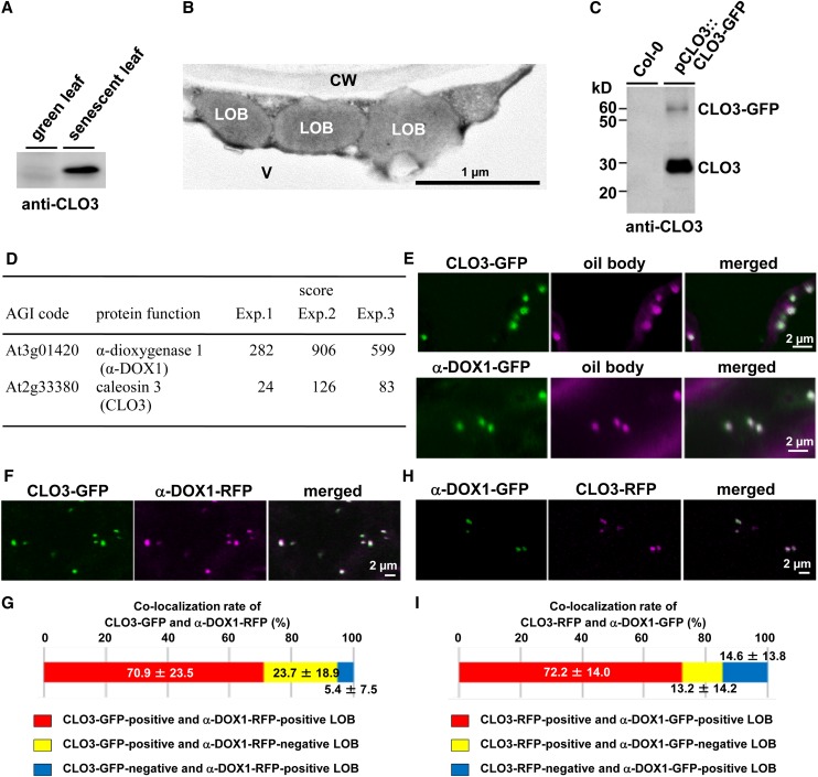
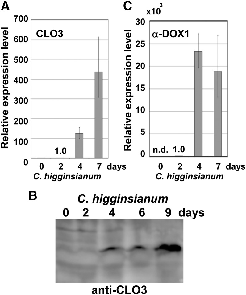
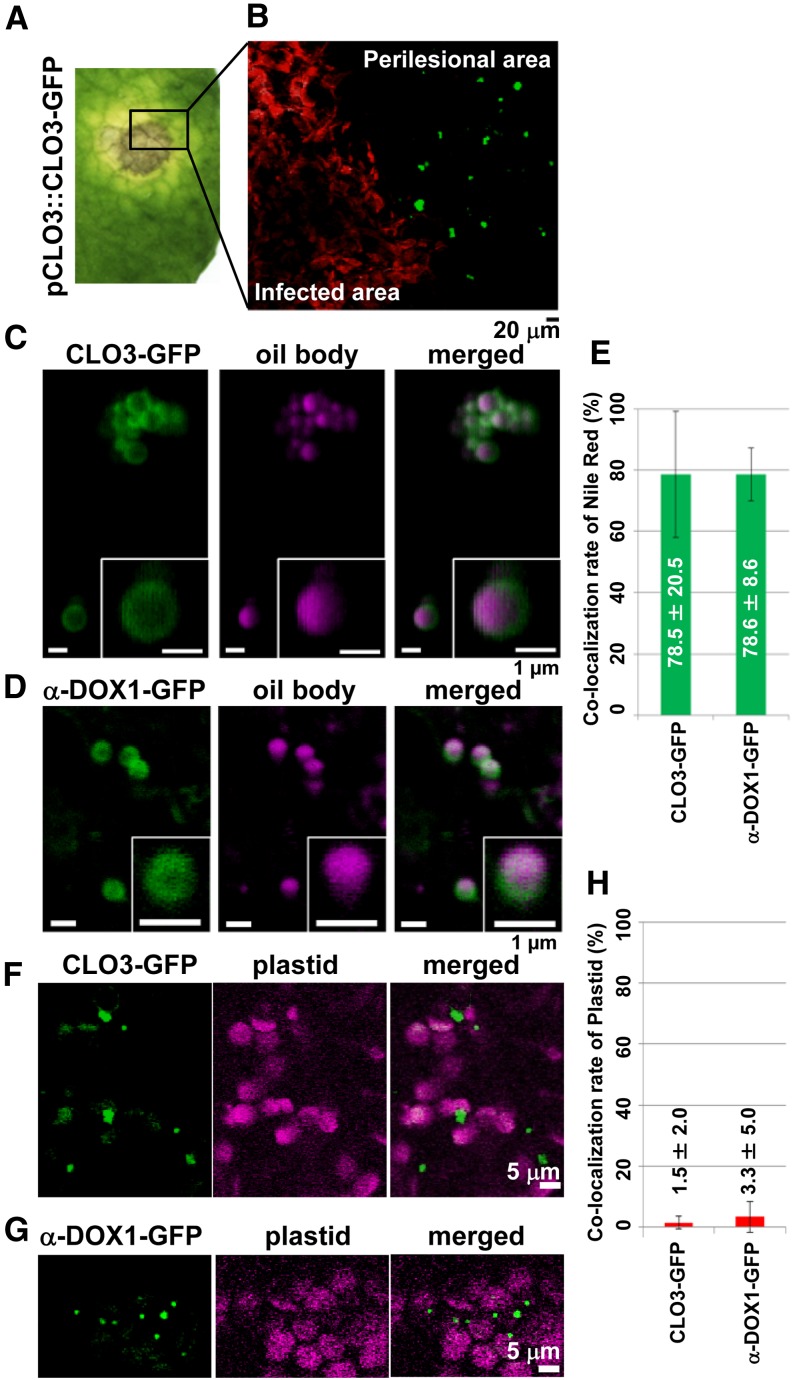
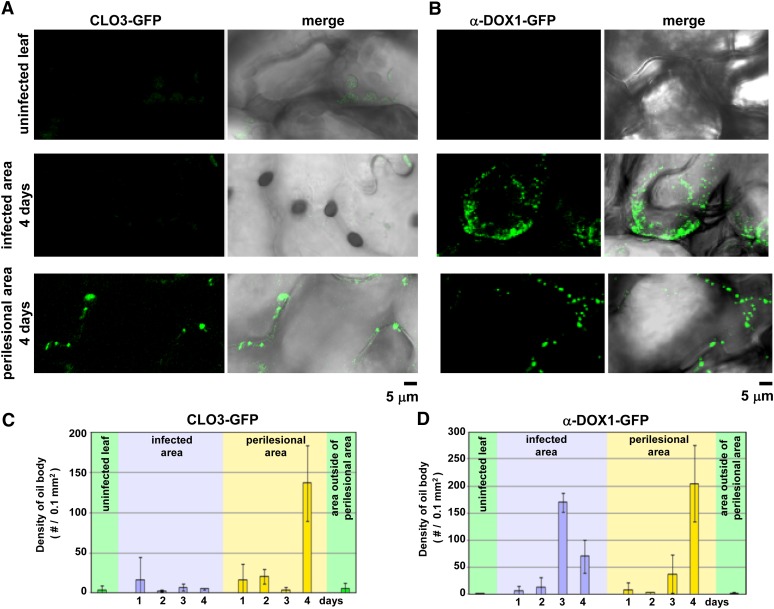
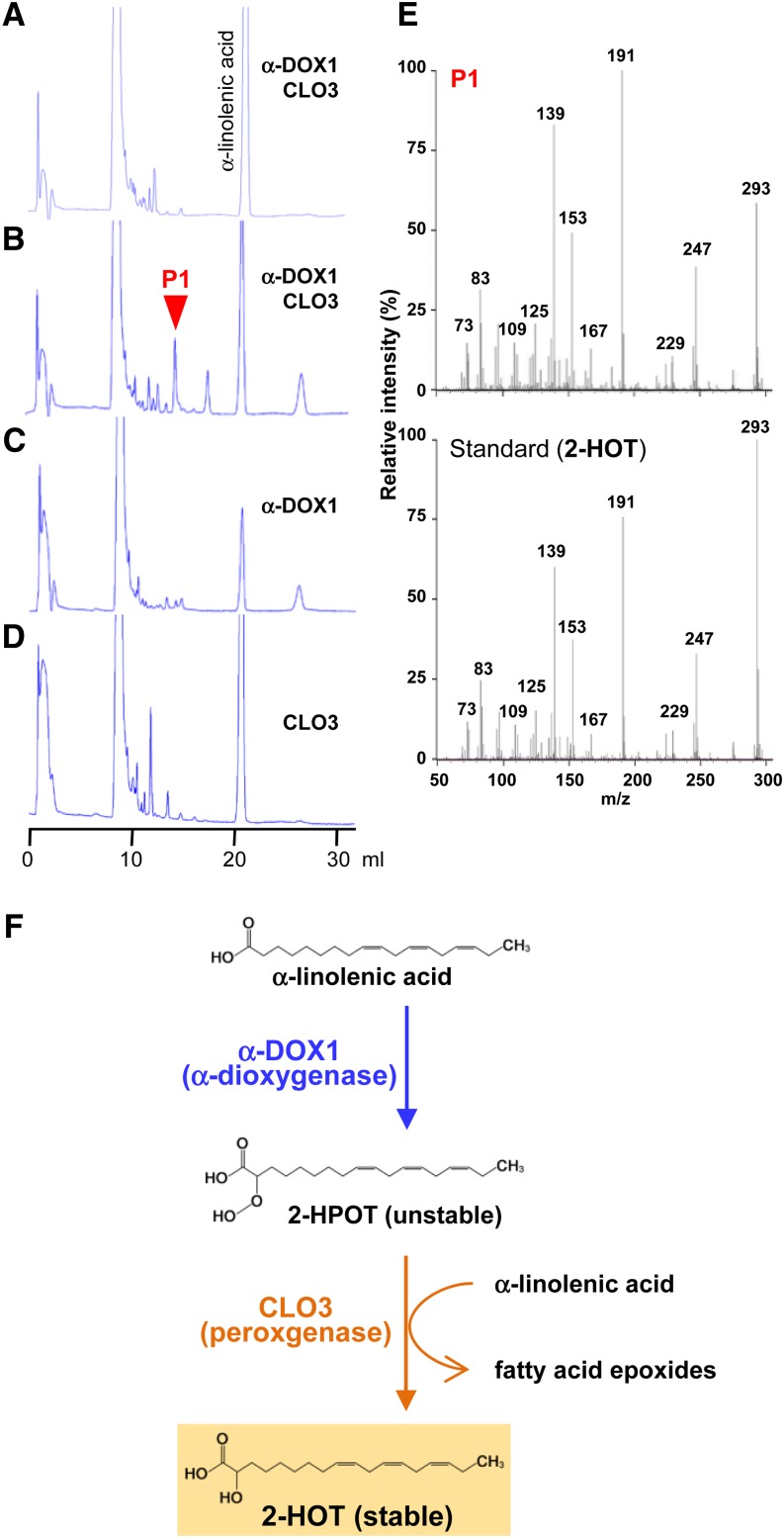
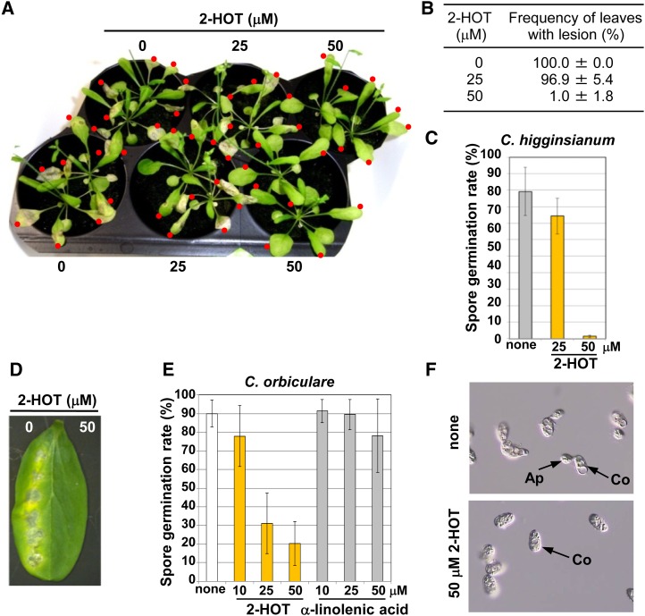
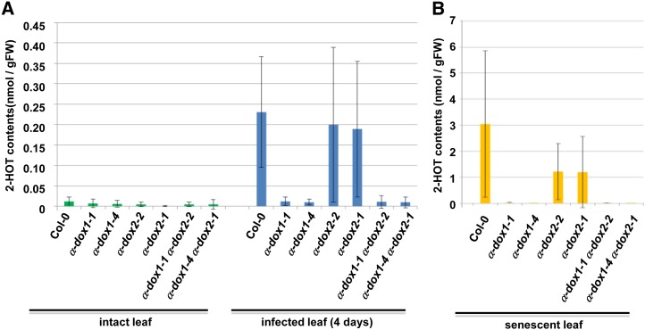
Oil bodies are intracellular structures present in the seed and leaf cells of many land plants. Seed oil bodies are known to function as storage compartments for lipids. However, the physiological function of leaf oil bodies is unknown. Here, we show that leaf oil bodies function as subcellular factories for the production of a stable phytoalexin in response to fungal infection and senescence. Proteomic analysis of oil bodies prepared from Arabidopsis (Arabidopsis thaliana) leaves identified caleosin (CLO3) and α-dioxygenase (α-DOX1). Both CLO3 and α-DOX1 were localized on the surface of oil bodies. Infection with the pathogenic fungus Colletotrichum higginsianum promoted the formation of CLO3- and α-DOX1-positive oil bodies in perilesional areas surrounding the site of infection. α-DOX1 catalyzes the reaction from α-linolenic acid (a major fatty acid component of oil bodies) to an unstable compound, 2-hydroperoxy-octadecatrienoic acid (2-HPOT). Intriguingly, a combination of α-DOX1 and CLO3 produced a stable compound, 2-hydroxy-octadecatrienoic acid (2-HOT), from α-linolenic acid. This suggests that the colocalization of α-DOX1 and CLO3 on oil bodies might prevent the degradation of unstable 2-HPOT by efficiently converting 2-HPOT into the stable compound 2-HOT. We found that 2-HOT had antifungal activity against members of the genus Colletotrichum and that infection with C. higginsianum induced 2-HOT production. These results defined 2-HOT as an Arabidopsis phytoalexin. This study provides, to our knowledge, the first evidence that leaf oil bodies produce a phytoalexin under a pathological condition, which suggests a new mechanism of plant defense.


Figures

Figure 1.
Figure 1.
Colocalization of CLO3 and α-DOX1 on the leaf oil bodies. A, Immunoblots showing the induction of CLO3 by senescence. B, Electron micrographs of senescent leaves showing spherical leaf oil bodies in cytoplasm. CW, Cell wall; LOB, leaf oil body; V, vacuole. C, Immunoblots showing that the pull-down CLO3-GFP-tagged oil bodies contained both CLO3-GFP and endogenous CLO3. D, LTQ-Orbitrap MS of CLO3-GFP-tagged oil bodies identified CLO3 and α-DOX1. Arabidopsis Genome Initiative (AGI) codes and annotations are from The Arabidopsis Information Resource database (http://www.arabidopsis.org). Scores were calculated using the Mascot program (Matrix Science). Raw data are given in Supplemental Data S1. E, Localization of CLO3 and α-DOX1 to oil bodies stained with Nile red. Either CLO3-GFP or α-DOX1-GFP was transiently expressed in N. benthamiana leaves. F, Colocalization of CLO3-GFP and α-DOX1-RFP by transient expression assay in N. benthamiana leaves. G, Quantitative analysis of the colocalization rate of CLO3-GFP and α-DOX1-RFP in seven fluorescence images. A total of 229 oil bodies were examined, most of which (yellow) were CLO3-GFP- and α-DOX1-RFP-positive oil bodies. Data represent average values ± sd. H, Colocalization of CLO3-RFP and α-DOX1-GFP by transient expression assay in N. benthamiana leaves. I, Quantitative analysis of the colocalization rate of CLO3-RFP and α-DOX1-GFP in nine fluorescence images. A total of 188 oil bodies were examined, most of which (yellow) were CLO3-RFP- and α-DOX1-GFP-positive oil bodies. Data represent average values ± sd.
Figure 2.
Figure 2.
CLO3 and α-DOX1 are induced remarkably after infection with C. higginsianum. A, mRNA levels of CLO3 in whole leaves after fungal infection. The mRNA level at 2 d was defined as 1.0. Vertical bars indicate the 95% confidence intervals for three experiments. The ACTIN2 gene was included as a control. B, Changes in the levels of the CLO3 protein in response to infection with C. higginsianum. Extracts from leaves infected with C. higginsianum were subjected to immunoblotting with a specific antibody against CLO3. C, mRNA levels of α-DOX1 in whole leaves after fungal infection. The mRNA level at 2 d was defined as 1.0. Vertical bars indicate the 95% confidence intervals for three experiments. The ACTIN2 gene was included as a control. n.d., Not detected.
Figure 3.
Figure 3.
CLO3 and α-DOX1 localize to leaf oil bodies of the perilesional area after infection with C. higginsianum. A, Leaves of transgenic plants expressing CLO3-GFP under the control of the endogenous CLO3 promoter were infected with C. higginsianum. The yellow perilesional area surrounds the infected area 4 d after infection. B, Fluorescence image of a section of the infected leaf in A showing that the detection of CLO3-GFP was limited to the perilesional area (an area free of fungi) and was not observed in areas where RFP-labeled fungi proliferated. C, Localization of CLO3-GFP on the surface of oil bodies stained with Nile red in the perilesional area of a 4-d-infected leaf. Insets show magnified views. D, Localization of α-DOX1-GFP on the surface of oil bodies stained with Nile red in the perilesional area of a 6-d-infected leaf of a transgenic plant expressing α-DOX1-GFP under the control of the endogenous α-DOX1 promoter. Insets show magnified views. E, Quantitative analysis of the rate of oil bodies labeled with CLO3-GFP (C) or α-DOX1-GFP (D). A total of 73 oil bodies in three fluorescence images of CLO3-GFP and a total of 101 oil bodies in five fluorescence images of α-DOX1-GFP were examined in 4-d-infected leaves. Data represent average values ± sd (error bars). F and G, Autofluorescence images of plastids and GFP fluorescence images of CLO3-GFP (F) and α-DOX1-GFP (G) in the perilesional area of 4-d-infected leaves. H, Quantitative analysis of the rate of CLO3-GFP-positive dots (F) or α-DOX1-GFP-positive dots (G) overlapped with autofluorescence images of plastids. A total of 135 CLO3-GFP-positive dots in five fluorescence images and a total of 210 α-DOX1-GFP-positive dots in five fluorescence images were examined. Data represent average values ± sd (error bars).
Figure 4.
Figure 4.
Changes in the number of CLO3-GFP-positive and α-DOX1-GFP-positive oil bodies in the infected area and the yellow perilesional area. A, Punctate fluorescent structures in the leaves of pCLO3::CLO3-GFP transgenic plants 0, 2, and 4 d after infection with C. higginsianum. Uninfected leaves, the infected area, and the perilesional area were observed by confocal laser scanning microscopy (CLO3-GFP) and differential interference contrast (DIC). Right panels show merged images of CLO3-GFP and DIC. B, Punctate fluorescent structures in the leaves of pα-DOX1::α-DOX1-GFP transgenic plants 0, 2, and 4 d after infection with C. higginsianum. Uninfected leaves, the infected area, and the perilesional area were observed by confocal laser scanning microscopy (α-DOX1-GFP) and DIC. Right panels show merged images of α-DOX1-GFP and DIC. C and D, Changes in the density of CLO3-GFP-positive (C) and α-DOX1-positive (D) oil bodies (number of oil bodies per 0.1 mm2 of tissue) in the respective leaf area of the transgenic plants after infection. Vertical bars indicate sd of three individual leaves.
Figure 5.
Figure 5.
Recombinant proteins of CLO3 and α-DOX1 act cooperatively to catalyze the conversion of α-linolenic acid into 2-HOT. A to D, HPLC patterns of the products of the reaction of α-linolenic acid with α-DOX1 and CLO3 for 0 h (A) and 24 h (B) and with either α-DOX1 (C) or CLO3 (D) for 24 h. α-DOX1 and CLO3 together generate a new peak (P1) from α-linolenic acid. E, MS/MS profiles of the P1 fraction (top) and a standard of 2-HOT (bottom) identifying the product of the P1 fraction as 2-HOT. F, Possible α-dioxygenase pathway for the production of 2-HOT from α-linolenic acid involving the cooperative action of α-DOX1 and CLO3.
Figure 6.
Figure 6.
Antifungal activity of 2-HOT against C. higginsianum and C. orbiculare. A, Arabidopsis leaves that were drop inoculated with spore suspensions of C. higginsianum containing 2-HOT (0, 25, and 50 µm). Images were obtained 7 d after inoculation. Red dots indicate drop-inoculated leaves. B, Number of leaves forming lesions after infection with C. higginsianum. Statistical analyses were performed on three independent infection experiments. Photographs of the infected plants are shown in A and Supplemental Figure S5. C, Effect of 2-HOT on the germination rate of C. higginsianum spores on Arabidopsis leaves. Drops containing spores and the indicated compounds were placed on the leaves and viewed after 1 d. none, Solvent only (0.3% ethanol). Vertical bars indicate sd of three repeated experiments. D, Cucumber cotyledons were drop inoculated with spore suspensions of another species of Colletotrichum, C. orbiculare, containing 2-HOT (0, 25, and 50 µm). Images were obtained 7 d after the inoculation. E, Effect of 2-HOT on the germination rate of C. orbiculare spores. Drops containing spores and the indicated compounds were placed on a microscope slide and viewed after 3 h. none, Solvent only (0.3% ethanol). Vertical bars indicate sd of three repeated experiments. F, Photographs of the germination rate of C. orbiculare spores. Spore suspensions with or without 2-HOT-containing spores and the indicated compounds were placed on a microscope slide and viewed after 3 h. Ap, Appressorium; Co, conidia; none, spore suspension (0.3% ethanol).
Figure 7.
Figure 7.
The accumulation of 2-HOT in infected and senescent leaves is dependent on α-dioxygenase. 2-HOT contents in the wild type and the α-dioxygenase mutants (α-dox1-1, α-dox1-4, α-dox2-2, α-dox2-1, α-dox1-1 α-dox2-2, and α-dox1-4 α-dox2-1) were measured by LC-MS. Leaves at 4 d after C. higginsianum infection (A; infected leaf), senescent leaves (B), and intact leaves were used. Data represent average values ± sd (error bars). Details are provided in Supplemental Figure S6A. FW, Fresh weight.

Similar articles

Cited by

References

    1. Abell BM, Holbrook LA, Abenes M, Murphy DJ, Hills MJ, Moloney MM. (1997) Role of the proline knot motif in oleosin endoplasmic reticulum topology and oil body targeting. Plant Cell 9: 1481–1493 - PMC - PubMed
    1. Adie BA, Pérez-Pérez J, Pérez-Pérez MM, Godoy M, Sánchez-Serrano JJ, Schmelz EA, Solano R. (2007) ABA is an essential signal for plant resistance to pathogens affecting JA biosynthesis and the activation of defenses in Arabidopsis. Plant Cell 19: 1665–1681 - PMC - PubMed
    1. Ahuja I, Kissen R, Bones AM. (2012) Phytoalexins in defense against pathogens. Trends Plant Sci 17: 73–90 - PubMed
    1. Asakura M, Ninomiya S, Sugimoto M, Oku M, Yamashita S, Okuno T, Sakai Y, Takano Y. (2009) Atg26-mediated pexophagy is required for host invasion by the plant pathogenic fungus Colletotrichum orbiculare. Plant Cell 21: 1291–1304 - PMC - PubMed
    1. Aubert Y, Vile D, Pervent M, Aldon D, Ranty B, Simonneau T, Vavasseur A, Galaud JP. (2010) RD20, a stress-inducible caleosin, participates in stomatal control, transpiration and drought tolerance in Arabidopsis thaliana. Plant Cell Physiol 51: 1975–1987 - PubMed

Publication types

MeSH terms