

doi: 10.1093/nar/gkt1047.
Epub 2013 Nov 7.
The eukaryotic linear motif resource ELM: 10 years and counting
Affiliations
- PMID: 24214962
- PMCID: PMC3964949
- DOI: 10.1093/nar/gkt1047
Item in Clipboard
The eukaryotic linear motif resource ELM: 10 years and counting
Nucleic Acids Res.
2014 Jan.
Abstract
The eukaryotic linear motif (ELM http://elm.eu.org) resource is a hub for collecting, classifying and curating information about short linear motifs (SLiMs). For >10 years, this resource has provided the scientific community with a freely accessible guide to the biology and function of linear motifs. The current version of ELM contains ∼200 different motif classes with over 2400 experimentally validated instances manually curated from >2000 scientific publications. Furthermore, detailed information about motif-mediated interactions has been annotated and made available in standard exchange formats. Where appropriate, links are provided to resources such as switches.elm.eu.org and KEGG pathways.
Figures
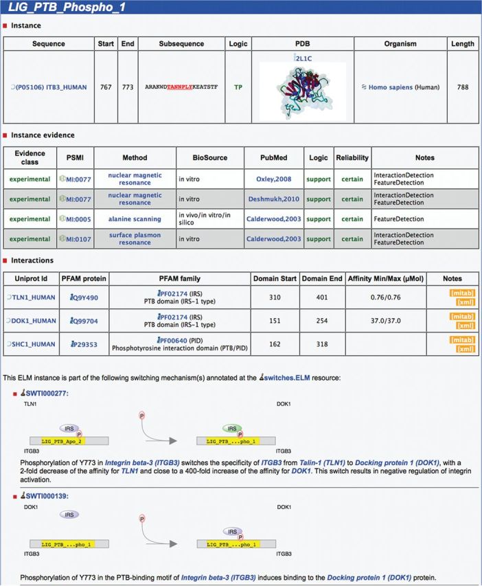
Screenshot of the ELM website showing details for an instance of the ELM class LIG_PTB_Phospho_1 in the human protein Integrin beta-3 at position 767–773. Details about the instance are depicted on top, including a representation of the 3D structure PDB:2LIC showing the instance bound by ‘SHC-transforming protein 1’. Below the instance evidence, which holds details about the methods used in the article, information regarding the interaction between the linear motif and the domain can be found. Here, three interaction partners containing phosphotyrosine-binding domains (PTB) are annotated: ‘talin-1’, ‘docking protein 1’ and ‘SHC-transforming protein 1’. Finally, the two schematics at the bottom illustrate the involvement of this motif instance in molecular switching mechanisms.
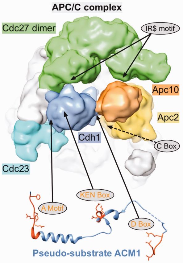
Motif-mediated regulation of APC/C function. Structure of the yeast APC/C complex [EMD-1815, determined by Cryo-EM. Figure generated with chimera (30)] with confirmed (full arrows) or putative (dashed arrows) motif-binding sites indicated. Binding of the co-activator Cdh1 (blue) to the APC/C is mediated by two motifs: The C-terminal IR motif binds to the tetratricopeptide repeat (TPR) region of one subunit of the Cdc27 dimer (green) and the C-Box binds to the catalytic Apc2 subunit (yellow). The Apc10 (orange) subunit also contains a C-terminal IR motif, which binds to the TPR domain of the second Cdc27 subunit (green). Recruitment of substrates or additional regulators such as the pseudo-substrate ACM1 (PDB:4BH6) also depends on motifs. The A Motif, KEN-Box and D-Box bind to different sites on the WD40 domain of Cdh1. In addition, the D-Box also contacts Apc10, which functions as a co-receptor for this degron (31).
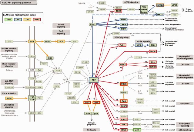
Motif-mediated interactions annotated in the ELM resource mapped onto the KEGG (40) human Phosphatidylinositol-3′-kinase-(PI3K)–Akt signaling pathway (hsa04151). The direction of arrows denotes pathway direction. A colored border around a protein name indicates a motif within this protein is responsible for mediating the interaction to another protein in this pathway, also highlighted by a colored edge—docking motifs (blue), degrons (green), ligand binding motifs (orange) and modification sites (red). Colored dotted lines represent motif-mediated interactions mapped by homology. Phosphorylation/dephosphorylation events are indicated as ‘+p’/‘−p’ next to a node, respectively.
References
-
- Tompa P. Intrinsically disordered proteins: a 10-year recap. Trends Biochem. Sci. 2012;37:509–516. - PubMed
-
- Dunker AK, Lawson JD, Brown CJ, Williams RM, Romero P, Oh JS, Oldfield CJ, Campen AM, Ratliff CM, Hipps KW, et al. Intrinsically disordered protein. J. Mol. Graph. Model. 2001;19:26–59. - PubMed
-
- Uversky VN, Gillespie JR, Fink AL. Why are “natively unfolded” proteins unstructured under physiologic conditions? Proteins. 2000;41:415–427. - PubMed
-
- Wright PE, Dyson HJ. Intrinsically unstructured proteins: re-assessing the protein structure-function paradigm. J. Mol. Biol. 1999;293:321–331. - PubMed
-
- Fuxreiter M, Tompa P, Simon I. Local structural disorder imparts plasticity on linear motifs. Bioinformatics. 2007;23:950–956. - PubMed
Publication types
MeSH terms
Substances
Grants and funding
LinkOut - more resources
Full Text Sources
Other Literature Sources
