. 2014 Feb;42(3):2037-48.
doi: 10.1093/nar/gkt1050. Epub 2013 Nov 7.

The mechanism of pseudouridine synthases from a covalent complex with RNA, and alternate specificity for U2605 versus U2604 between close homologs

Affiliations

The mechanism of pseudouridine synthases from a covalent complex with RNA, and alternate specificity for U2605 versus U2604 between close homologs

Nadine Czudnochowski et al. Nucleic Acids Res. 2014 Feb.

Abstract

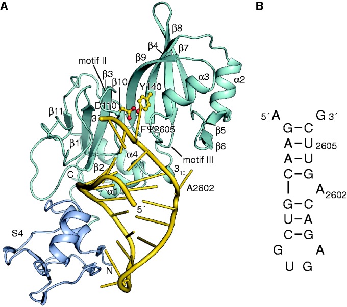
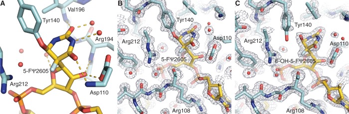
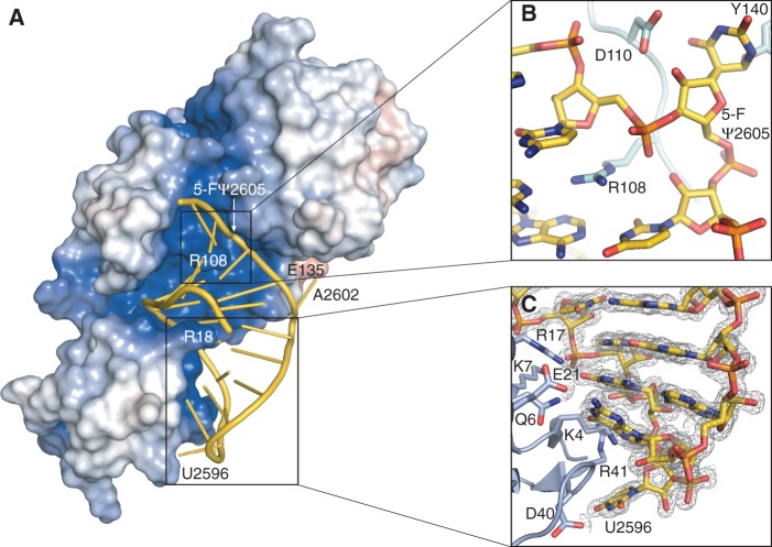
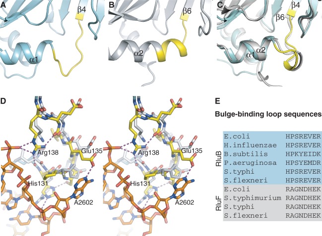
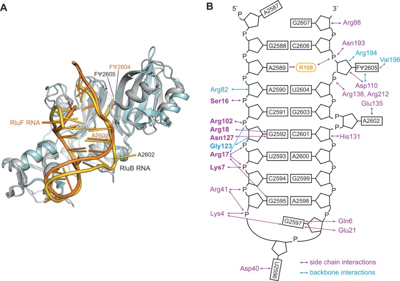
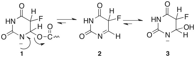
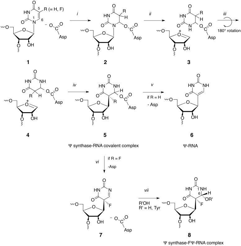
RluB catalyses the modification of U2605 to pseudouridine (Ψ) in a stem-loop at the peptidyl transferase center of Escherichia coli 23S rRNA. The homolog RluF is specific to the adjacent nucleotide in the stem, U2604. The 1.3 Å resolution crystal structure of the complex between the catalytic domain of RluB and the isolated substrate stem-loop, in which the target uridine is substituted by 5-fluorouridine (5-FU), reveals a covalent bond between the isomerized target base and tyrosine 140. The structure is compared with the catalytic domain alone determined at 2.5 Å resolution. The RluB-bound stem-loop has essentially the same secondary structure as in the ribosome, with a bulge at A2602, but with 5-FU2605 flipped into the active site. We showed earlier that RluF induced a frame-shift of the RNA, moving A2602 into the stem and translating its target, U2604, into the active site. A hydrogen-bonding network stabilizes the bulge in the RluB-RNA but is not conserved in RluF and so RluF cannot stabilize the bulge. On the basis of the covalent bond between enzyme and isomerized 5-FU we propose a Michael addition mechanism for pseudouridine formation that is consistent with all experimental data.


Figures

Figure 1.
Figure 1.
Overall structure of the RluB-RNA complex. (A) Structure of RluB-bound to a small RNA substrate. The S4 domain is blue, the catalytic domain is blue–green and the RNA phosphate backbone is gold. Nucleosides are shown as gold sticks. The catalytic aspartate 110 and tyrosine 140 are shown in ball-and-stick form with side chains colored gold. (B) Schematic drawing of the 21-mer rRNA small substrate of RluB. For crystallization U2605 was substituted by 5-FU.
Figure 2.
Figure 2.
Active site of the RluB-RNA complex. (A) View of the active site of the first molecule with protein and RNA shown as sticks and H-bonding interactions of the target base and ribose with neighboring protein residues and water depicted as gold dashes. (B) and (C) 2Fo-Fc (αcalc) density at the active sites of the two molecules in the asymmetric unit of RluB–RNA crystals, contoured at 1.5 σ and 1.2 σ, respectively. (B) The density map for the first molecule clearly shows a covalent bond between conserved Tyr140 and the isomerized target base. Asp110 is the catalytic Asp. (C) Tyr140 in the second molecule is in partial density, indicating this molecule may exist as a mixture of covalent and non-covalent RNA complexes in the crystal.
Figure 3.
Figure 3.
Interactions between RluB and the 21-mer RNA stem-loop. (A) Surface representation of RluB colored according to its electrostatic surface potential with positive electrostatic potential in blue (+7 kT/e) and negative potential in red (–7 kT/e). The 21-mer RNA is represented in gold. The orientation of RluB is as in Figure 1A. (B) Arginine 108 intercalates into the RNA stem. RNA and protein residues are shown as gold and blue–green sticks, respectively. (C) The S4 domain of RluB anchors the loop of the RNA to the protein. RluB is shown in blue, side chains interacting with the RNA (colored gold) are depicted as sticks. The 2Fo-Fc density for the RNA is contoured at 1.5 σ.
Figure 4.
Figure 4.
Comparison of the bulge-binding loop in RluB and RluF. (A) apo-RluB is colored blue–green and the bulge-binding loop (residues 131–138) is colored yellow. (B) apo-RluF in gray with analogous residues (128–135) colored yellow. (C) Superposed RluB and RluF in their RNA-bound conformations (RNA omitted for clarity). RluB is blue–green with its bulge-binding loop highlighted in yellow. RluF is gray. The loops have refolded to the same structure in the two RNA-bound complexes. (D) Stereo plot of the superposed RluB– and RluF–RNA complexes in the vicinity of the bulge (A2602). RluB is plotted with blue–green carbons and RluF with gray carbons. The bulge-binding loop in RluB is shown with yellow carbons and hydrogen bonds from this loop are shown as purple-dashed lines. (E) Aligned sequences of loop residues from several species of RluB and RluF.
Figure 5.
Figure 5.
RNA recognition by RluB and RluF. (A) Comparison of RluB–RNA (blue–green and gold) that targets U2605 and RluF–RNA (gray and orange) that targets U2604. In the latter A2602 is refolded into the stem, base registry is shifted up by one base on the site 3′ of A2602, and U2604 is flipped out (11). The RNAs overlay closely in the RNA-binding grooves of the catalytic domains. (B) Representation of hydrogen bond interactions between residues of RluB and nucleotides of the RNA stem-loop. Residues that are conserved in RluF and engage in interactions with the same nucleotide (from A2590 to U2596) as RluB are highlighted in bold.
Scheme 3.
Scheme 3.
Conversion of 5-F-6-acyloxy-uracil to 5-F-6-hydroxy-uracil.
Scheme 4.
Scheme 4.
Complete current mechanism of Ψ synthase and its reaction with 5-FU-RNA.

Similar articles

Cited by

References

    1. Bjork G. Stable RNA Modification. In: Neidhardt FC, Curtiss R, editors. Escherichia coli and Salmonella: Cellular and Molecular Biology. 2nd edn. Washington, DC: ASM Press; 1996. pp. 861–886.
    1. Anantharaman V, Koonin EV, Aravind L. Comparative genomics and evolution of proteins involved in RNA metabolism. Nucleic Acids Res. 2002;30:1427–1464. - PMC - PubMed
    1. Moore PB, Steitz TA. The involvement of RNA in ribosome function. Nature. 2002;418:229–235. - PubMed
    1. Ofengand J. Ribosomal RNA pseudouridines and pseudouridine synthases. FEBS Lett. 2002;514:17–25. - PubMed
    1. Huang L, Pookanjanatavip M, Gu X, Santi DV. A conserved aspartate of tRNA pseudouridine synthase is essential for activity and a probable nucleophilic catalyst. Biochemistry. 1998;37:344–351. - PubMed

Publication types

MeSH terms

LinkOut - more resources