. 2013 Dec 19;122(26):4210-9.
doi: 10.1182/blood-2012-12-472803. Epub 2013 Nov 8.

Human proT-cells generated in vitro facilitate hematopoietic stem cell-derived T-lymphopoiesis in vivo and restore thymic architecture

Affiliations

Human proT-cells generated in vitro facilitate hematopoietic stem cell-derived T-lymphopoiesis in vivo and restore thymic architecture

Génève Awong et al. Blood. .

Abstract

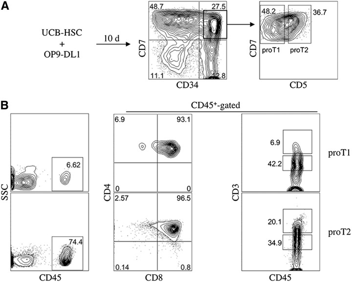
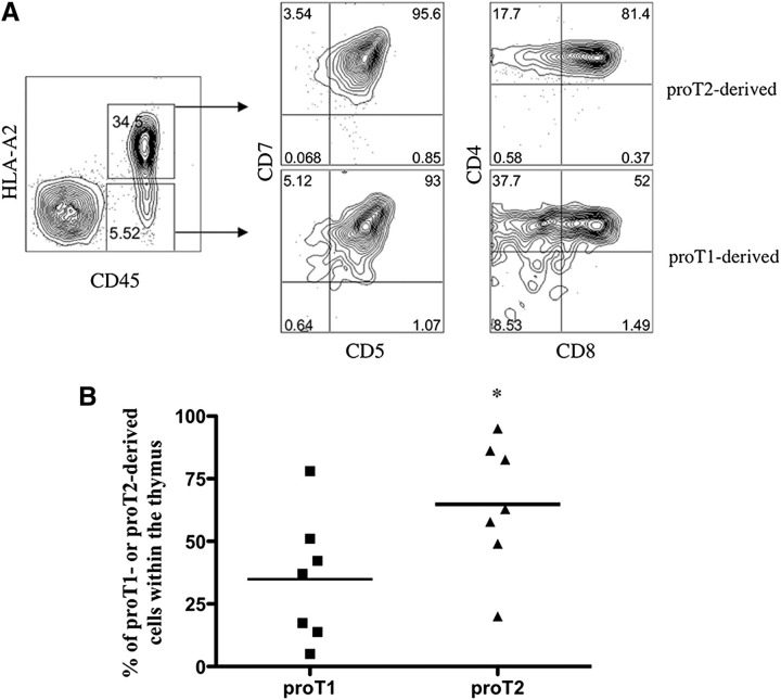
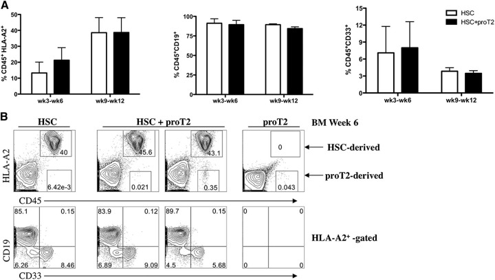
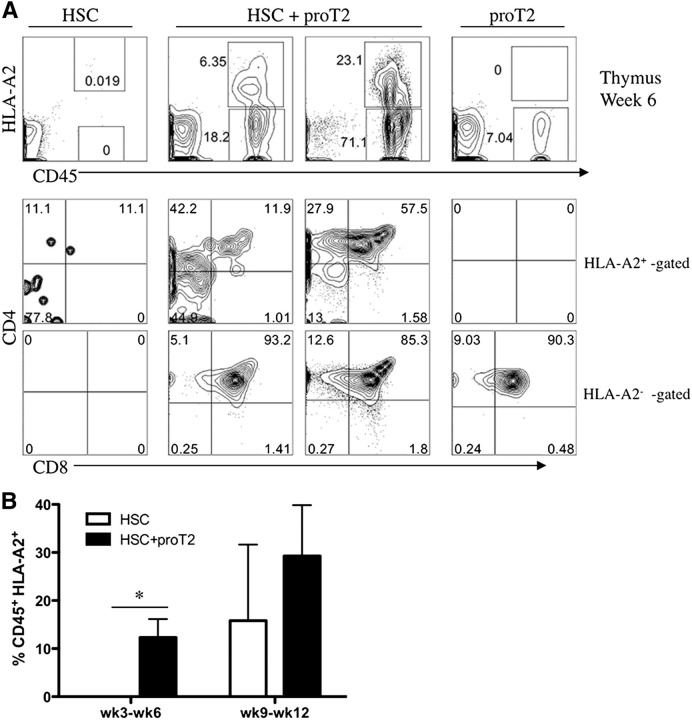
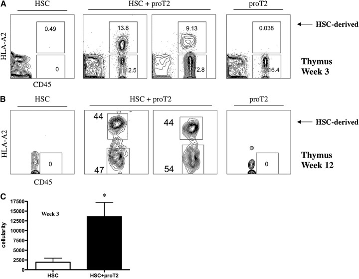
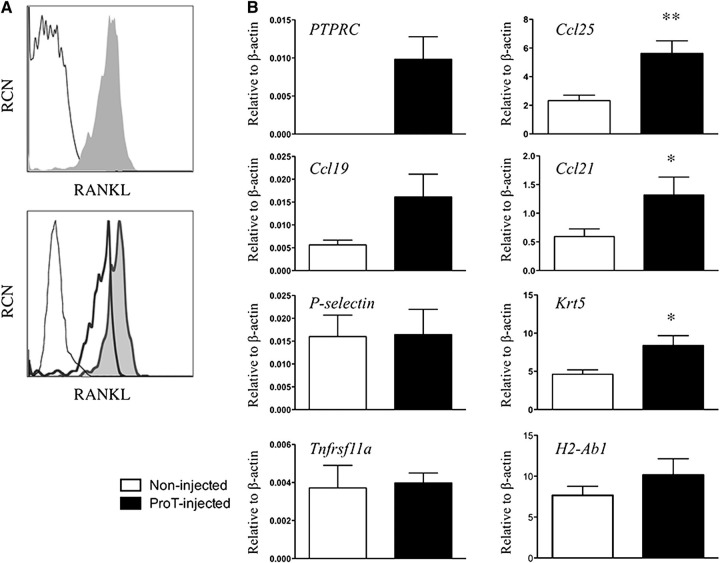
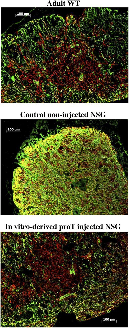
Hematopoietic stem cell transplantation (HSCT) is followed by a period of immune deficiency due to a paucity in T-cell reconstitution. Underlying causes are a severely dysfunctional thymus and an impaired production of thymus-seeding progenitors in the host. Here, we addressed whether in vitro-derived human progenitor T (proT)-cells could not only represent a source of thymus-seeding progenitors, but also able to influence the recovery of the thymic microenvironment. We examined whether co-transplantation of in vitro-derived human proT-cells with hematopoietic stem cells (HSCs) was able to facilitate HSC-derived T-lymphopoiesis posttransplant. A competitive transfer approach was used to define the optimal proT subset capable of reconstituting immunodeficient mice. Although the 2 subsets tested (proT1, CD34(+)CD7(+)CD5(-); proT2, CD34(+)CD7(+)CD5(+)) showed thymus engrafting function, proT2-cells exhibited superior engrafting capacity. Based on this, when proT2-cells were coinjected with HSCs, a significantly improved and accelerated HSC-derived T-lymphopoiesis was observed. Furthermore, we uncovered a potential mechanism by which receptor activator of nuclear factor κb (RANK) ligand-expressing proT2-cells induce changes in both the function and architecture of the thymus microenvironment, which favors the recruitment of bone marrow-derived lymphoid progenitors. Our findings provide further support for the use of Notch-expanded progenitors in cell-based therapies to aid in the recovery of T-cells in patients undergoing HSCT.


Figures

Figure 1
Figure 1
Flow cytometric analysis of engraftment and differentiation of in vitroderived CD34+ CD7++ CD5 (proT1)- and CD34+ CD7++ CD5+ (proT2)-cells in immunodeficient mice. (A) Human UCB CD34+CD38−/lo cells were differentiated on OP9-DL1 cells for 10 days and CD34+CD7++ CD5 (proT1)- and CD34+ CD7++ CD5+ (proT2)-cells were sorted by flow cytometry. (B) Neonatal NSG mice were injected intrahepatically with 2.0 × 10⁵ cells of either subset (n ≥ 8 mice/group). Thymuses were harvested after 4 weeks and stained for CD45, CD4, CD8, and CD3. The percentage of human CD45+ cells present in the thymus of NSG mice transplanted with proT1-cells (average thymus cellularity 27.6 × 10⁴ ± 6.6 × 10⁴) or proT2 cells (average thymus cellularity 31.6 × 10⁴ ± 4.0 × 10⁴) are shown. CD4, CD8, and CD3 cell surface expressions are shown on CD45+-gated thymocytes. The results shown are representative of at least 3 independent experiments. SSC, side scatter.
Figure 2
Figure 2
Analysis for the presence of proT1-derived and proT2-derived cells in the thymus of competitively reconstituted immunodeficient mice. (A) 1:1 mixture of sorted HLA-A2 proT1-cells (1 × 10⁵) and sorted HLA-A2+ (1 × 10⁵) proT2-cells were injected into nonirradiated NSG neonatal mice and the thymuses harvested and analyzed after 17 days (n = 7 mice/group). Shown is flow cytometric analysis of human CD45 and HLA-A2 cell surface expression and CD5, CD7, CD8, and CD4 expression on CD45+HLA-A2– and CD45+HLA-A2+–gated cells for proT1- or proT2-derived cells, respectively. (B) Percentage of HLA-A2 or HLA-A2+ cells over total human CD45+ cells for individual mice shown. The results shown are representative of at least 3 independent experiments. *P < .05.
Figure 3
Figure 3
Analysis of engraftment and differentiation of HSCs and proT2-cells in the BM of immunodeficient mice. (A) Percentage of human CD45+HLA-A2+ (left), CD19 (middle), and CD33 (right) HSC-derived engraftment at 3 to 6 weeks, and 9 to 12 weeks after intrahepatic injection of neonatal NSG mice receiving either HSCs (white) or HSCs with proT2-cells (black). (B) Flow cytometric analysis of CD45 vs HLA-A2, and CD19 and CD33 on CD45+HLA-A2+–gated cells in the BM at 6 weeks postintrahepatic injection. Table 2 shows the number of mice analyzed. The results shown are representative of at least 3 independent experiments.
Figure 4
Figure 4
Analysis of engraftment and differentiation of HSCs and proT2-cells in the thymus of immunodeficient mice. (A) Flow cytometric analysis of CD45 vs HLA-A2, and CD4 and CD8 on CD45+HLA-A2+–gated (HSC-derived) and CD45+HLA-A2 –gated (proT2-derived) cells in the thymus at 6 weeks after intrahepatic injection of NSG mice receiving either HSCs, HSCs with proT2-cells, or proT2-cells. (B) Percentage of human CD45+HLA-A2+ HSC-derived engraftment at 3 to 6 weeks and 9 to 12 weeks after intrahepatic injection of neonatal mice receiving either HSCs (white) or HSCs with proT2 cells (black). Table 2 shows the number of mice analyzed. The results shown are representative of at least 3 independent experiments. *P < .05.
Figure 5
Figure 5
Analysis of early and late engraftment, and differentiation of HSCs and proT2-cells in the thymus of immunodeficient mice. Flow cytometric analysis of CD45 vs HLA-A2 in the thymus at (A) 12 weeks and at (B) 3 weeks, postintrahepatic injection of neonatal NSG mice receiving HSCs, HSCs with proT2-cells, or proT2-cells (n = 5 and n = 12 mice/group, respectively). Human CD45 cells represent cells of host mouse origin. (C) Thymus cellularity in mice receiving HSCs or HSCs with proT2-cells 3 weeks after injection. The results shown are representative of at least 3 independent experiments. Cellularity was calculated based on human CD45+ cells obtained by flow cytometry. *P < .05.
Figure 6
Figure 6
Gene expression analyses from thymuses obtained from in vitro–derived proT-injected mice. (A) Flow cytometric analysis of RANK ligand (RANKL) expression on purified CD34+CD38 cells (not shaded) and day 11 in vitro–derived CD34+CD7+-gated proT-cells (shaded) (top); and in vitro–derived proT1- (not shaded, thick line) and proT2-cells (shaded) (bottom). Unstained cells are included as a control (not shaded, thin dashed line). (B) Quantitative real time reverse transcriptase polymerase chain reaction analysis for the expression of human PTPRC (CD45), and mouse Ccl25, Ccl19, Ccl21, Selp (P-selectin), Krt5 (Cytokeratin-5), Tnfrsf11a (RANK), and H2-Ab1 (MHC class II) from mouse thymus extracts of NSG mice injected with proT-cells or control noninjected mice after 3 weeks. Transcript levels for all genes were normalized to mouse β-actin. These results are the average of 3 independent experiments, with the exception of Selp (n = 2), with error bars corresponding to standard error of the mean. Asterisks represent statistical significance as determined by Student t tests. The results shown are representative of 3 independent experiments. *P < .05; **P < .005.
Figure 7
Figure 7
Immunohistologic analysis of thymus. Sections of the thymus from an adult WT mouse (top); control noninjected NSG mouse (middle); and in vitro–derived proT-injected NSG mouse (bottom) 6 weeks after intrahepatic injection into neonates (n = 6). Thymus tissues were stained with anti-Cytokeratin 8 (green; cortical) and anti-Cytokeratin 5 (red; medullary). The results shown are representative of 3 independent experiments.

References

    1. Holländer G, Gill J, Zuklys S, Iwanami N, Liu C, Takahama Y. Cellular and molecular events during early thymus development. Immunol Rev. 2006;209:28–46. - PubMed
    1. Blackburn CC, Manley NR. Developing a new paradigm for thymus organogenesis. Nat Rev Immunol. 2004;4(4):278–289. - PubMed
    1. Petrie HT, Zúñiga-Pflücker JC. Zoned out: functional mapping of stromal signaling microenvironments in the thymus. Annu Rev Immunol. 2007;25:649–679. - PubMed
    1. Shortman K, Wu L. Early T lymphocyte progenitors. Annu Rev Immunol. 1996;14:29–47. - PubMed
    1. Goldschneider I. Cyclical mobilization and gated importation of thymocyte progenitors in the adult mouse: evidence for a thymus-bone marrow feedback loop. Immunol Rev. 2006;209:58–75. - PubMed

Publication types

MeSH terms

Supplementary concepts