

In vivo confocal microscopy of the ocular surface: from bench to bedside
- PMID: 24215436
- PMCID: PMC3960287
- DOI: 10.3109/02713683.2013.842592
In vivo confocal microscopy of the ocular surface: from bench to bedside
Abstract
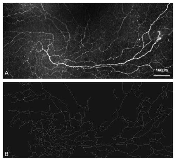
In vivo confocal microscopy (IVCM) is an emerging technology that provides minimally invasive, high resolution, steady-state assessment of the ocular surface at the cellular level. Several challenges still remain but, at present, IVCM may be considered a promising technique for clinical diagnosis and management. This mini-review summarizes some key findings in IVCM of the ocular surface, focusing on recent and promising attempts to move "from bench to bedside". IVCM allows prompt diagnosis, disease course follow-up, and management of potentially blinding atypical forms of infectious processes, such as acanthamoeba and fungal keratitis. This technology has improved our knowledge of corneal alterations and some of the processes that affect the visual outcome after lamellar keratoplasty and excimer keratorefractive surgery. In dry eye disease, IVCM has provided new information on the whole-ocular surface morphofunctional unit. It has also improved understanding of pathophysiologic mechanisms and helped in the assessment of prognosis and treatment. IVCM is particularly useful in the study of corneal nerves, enabling description of the morphology, density, and disease- or surgically induced alterations of nerves, particularly the subbasal nerve plexus. In glaucoma, IVCM constitutes an important aid to evaluate filtering blebs, to better understand the conjunctival wound healing process, and to assess corneal changes induced by topical antiglaucoma medications and their preservatives. IVCM has significantly enhanced our understanding of the ocular response to contact lens wear. It has provided new perspectives at a cellular level on a wide range of contact lens complications, revealing findings that were not previously possible to image in the living human eye. The final section of this mini-review provides a focus on advances in confocal microscopy imaging. These include 2D wide-field mapping, 3D reconstruction of the cornea and automated image analysis.
Conflict of interest statement
The authors report no conflicts of interest. The authors alone are responsible for the content and writing of this article.
Figures
References
-
- Niederer RL, McGhee CN. Clinical in vivo confocal microscopy of the human cornea in health and disease. Prog Retin Eye Res. 2010;29:30–58. - PubMed
-
- Guthoff RF, Zhivov A, Stachs O. In vivo confocal microscopy, an inner vision of the cornea – a major review. Clin Experiment Ophthalmol. 2009;37:100–117. - PubMed
-
- McLaren JW, Nau CB, Patel SV, Bourne WM. Measuring corneal thickness with the ConfoScan 4 and Z-ring adapter. Eye Contact Lens. 2007;33:185–190. - PubMed
-
- Thomas PA, Geraldine P. Infectious keratitis. CurrOpin Infect Dis. 2007;20:129–141. - PubMed
-
- Keay L, Edwards K, Naduvilath T, Taylor HR, Snibson GR, Forde K, et al. Microbial keratitis predisposing factors and morbidity. Ophthalmology. 2006;113:109–116. - PubMed
Publication types
MeSH terms
Grants and funding
LinkOut - more resources
Full Text Sources
Other Literature Sources
Medical