. 2013 Nov;123(11):4731-8.
doi: 10.1172/JCI67603.

NADPH oxidase 4 limits bone mass by promoting osteoclastogenesis

NADPH oxidase 4 limits bone mass by promoting osteoclastogenesis

Claudia Goettsch et al. J Clin Invest. 2013 Nov.

Abstract

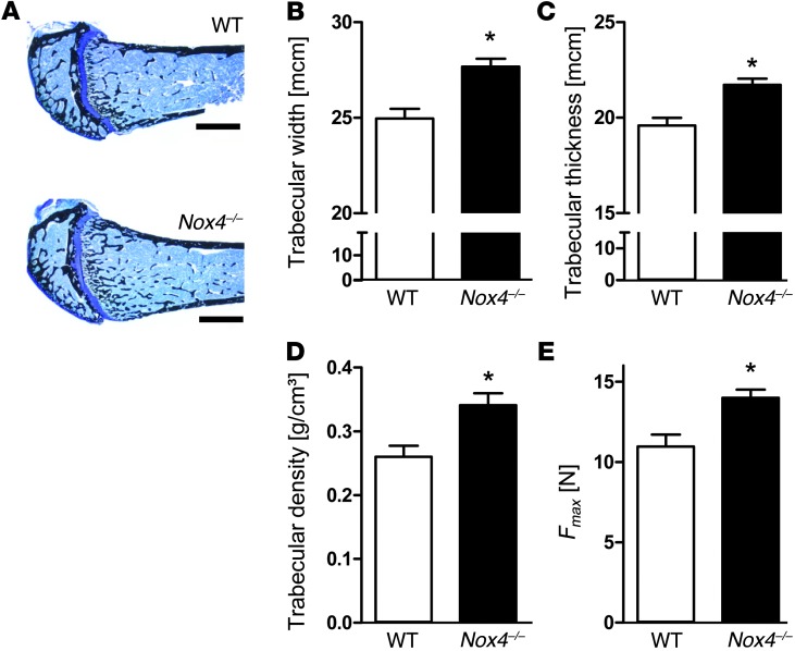
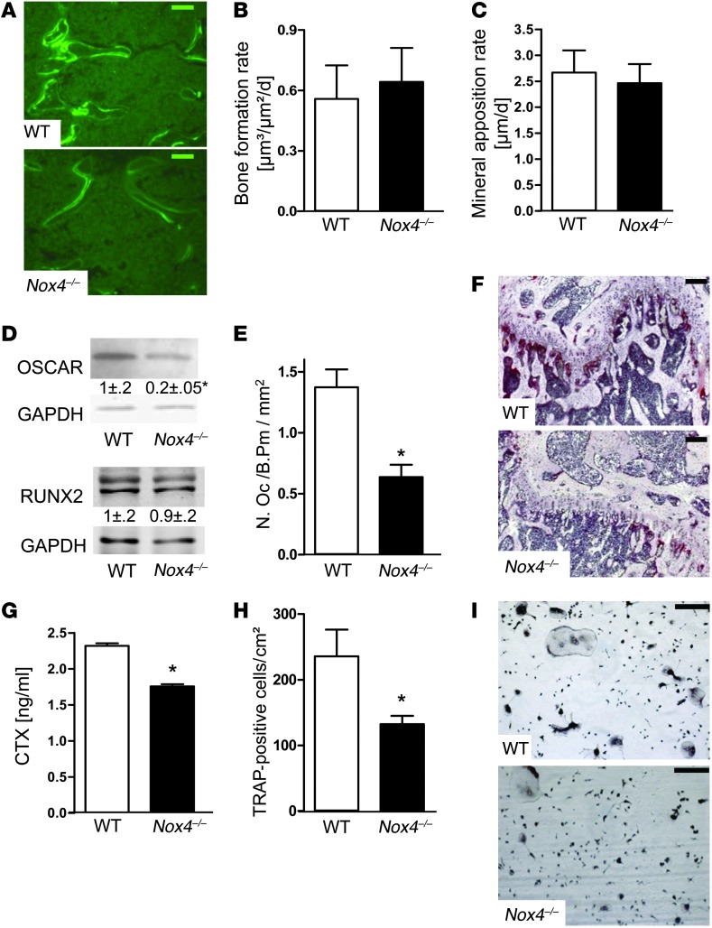
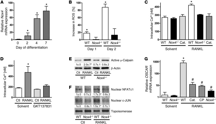
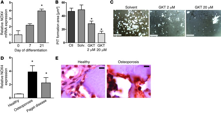
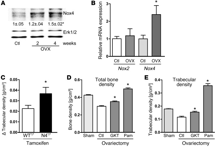
ROS are implicated in bone diseases. NADPH oxidase 4 (NOX4), a constitutively active enzymatic source of ROS, may contribute to the development of such disorders. Therefore, we studied the role of NOX4 in bone homeostasis. Nox4(-/-) mice displayed higher bone density and reduced numbers and markers of osteoclasts. Ex vivo, differentiation of monocytes into osteoclasts with RANKL and M-CSF induced Nox4 expression. Loss of NOX4 activity attenuated osteoclastogenesis, which was accompanied by impaired activation of RANKL-induced NFATc1 and c-JUN. In an in vivo model of murine ovariectomy–induced osteoporosis, pharmacological inhibition or acute genetic knockdown of Nox4 mitigated loss of trabecular bone. Human bone obtained from patients with increased osteoclast activity exhibited increased NOX4 expression. Moreover, a SNP of NOX4 was associated with elevated circulating markers of bone turnover and reduced bone density in women. Thus, NOX4 is involved in bone loss and represents a potential therapeutic target for the treatment of osteoporosis.


Figures

Figure 1
Figure 1. Effect of genetic knockout of Nox4 on bone density and morphometry.
(AC) Quantitative histomorphometry of the distal femur of WT and Nox4–/– mice (n = 3–5). (A) Representative undecalcified section of the distal femur stained with von Kossa. (B) Trabecular width. (C) Trabecular thickness. (D) Trabecular BMD, measured by peripheral quantitative computed tomography (n = 12). (E) Biomechanical properties of bone. Bone strength was tested by 3-point bending test (n = 9–11). Scale bars: 1 mm. Data are mean ± SEM. *P < 0.05.
Figure 2
Figure 2. Role of NOX4 in bone remodeling.
(A) Representative images of calcein-labeled bone surface. (B) Bone formation rate and (C) mineral apposition rate in bones of WT and Nox4–/– mice. (D) Western blot analysis and statistical analyses of densitometry (values shown relative to WT control) from femur lysates for OSCAR, RUNX2, and GAPDH as loading control. (E) Number of TRAP-positive osteoclasts (N.Oc) per bone perimeter (B.Pm), counted in histomorphometric analysis, and (F) representative images of TRAP-stained sections counterstained with Mayer’s hemalaun. (G) Serum levels of CTX. (H and I) Number (H) and representative images (I) of osteoclasts differentiated ex vivo from bone marrow cells of WT and Nox4–/– mice, as identified by TRAP staining and multinucleation. Scale bars: 500 μm (A); 100 μm (F and I). Data are mean ± SEM (n = 3–9). *P < 0.05.
Figure 3
Figure 3. Role of NOX4 in RANKL-induced signaling.
(A) Nox4 mRNA expression in murine predifferentiated BMNCs during osteoclastogenesis. (B) Relative change in ROS formation, as measured by Amplex Red/HRP, in mononuclear cells harvested from the spleen (splenocytes) stimulated with RANKL for 1 or 2 days. (C and D) Intracellular Ca2+, measured by Fura-2 fluorescence, in cells pretreated with (RANKL) or without RANKL (solvent) for 30 hours. Cells from WT and Nox4–/– mice were treated with and without (C) PEG-catalase (Cat.; 50 U/ml) or (D) GKT137831 (20 μM). (E and F) Representative Western blot and statistical analyses of densitometry (values shown relative to WT control) for the indicated proteins from WT and Nox4–/– cells with or without (ctl) RANKL prestimulation (50 ng/ml for 30 hours). (E) Total cellular lysates. (F) Nuclear fraction. (G) Relative mRNA expression of Oscar in cells treated with and without RANKL, PEG-catalase (50 U/ml), or calpeptin (CP; 20 μM) as indicated. Data are mean ± SEM (n = 5–8). *P < 0.05. #P < 0.05 vs. RANKL-treated WT.
Figure 4
Figure 4. Role of NOX4 in human bone loss.
(A) Nox4 mRNA expression in the course of differentiation of human precursor cells into osteoclasts. (B) Statistical analysis and (C) representative images of PIT formation assays of human PBMCs treated with RANKL with or without GKT137831. (D) Intensity of NOX4 staining in human bone material from healthy subjects and patients with osteoporosis or Paget disease. (E) Representative images of bone slides from a healthy subject and an osteoporotic patient stained for NOX4 (brown) and counterstained with hemalaun (violet). Scale bars: 1 mm (C); 50 μm (E). Data are mean ± SEM (n = 3–6). *P < 0.05.
Figure 5
Figure 5. Role of NOX4 for bone loss in the murine model of osteoporosis.
(A) Western blot analyses and statistical analyses of densitometry (values shown relative to healthy control) of NOX4 protein expression in bones of healthy and ovariectomized mice. (B) mRNA expression of Nox2 and Nox4 (n = 3–5). *P < 0.05. (CE) Bone density of the distal femur 6 weeks after ovariectomy. (C) Trabecular density increase in tamoxifen-treated WT (WT*/*) and Nox4fl/fl-ERT2-Cre+/0 (N4*/*) mice relative to ovariectomized, untreated WT animals (n = 8–12). *P < 0.05. (D) Total and (E) trabecular bone density, measured within 2 mm of the tibia plateau, in WT animals treated with solvent (Ctl), GKT137928 (GKT; 20 mg/kg/d), or pamidronate (Pam; 10 mg/kg once a week i.p.) beginning 2 days after ovariectomy (n = 8–15). Data are mean ± SEM. *P < 0.05 vs. solvent control.

Comment in

References

    1. Hsu H, et al. Tumor necrosis factor receptor family member RANK mediates osteoclast differentiation and activation induced by osteoprotegerin ligand. Proc Natl Acad Sci U S A. 1999;96(7):3540–3545. doi: 10.1073/pnas.96.7.3540. - DOI - PMC - PubMed
    1. Woo KM, Kim HM, Ko JS. Macrophage colony-stimulating factor promotes the survival of osteoclast precursors by up-regulating Bcl-X(L). Exp Mol Med. 2002;34(5):340–346. doi: 10.1038/emm.2002.48. - DOI - PubMed
    1. Kong YY, et al. OPGL is a key regulator of osteoclastogenesis, lymphocyte development and lymph-node organogenesis. Nature. 1999;397(6717):315–323. doi: 10.1038/16852. - DOI - PubMed
    1. Lacey DL, et al. Osteoprotegerin ligand modulates murine osteoclast survival in vitro and in vivo. Am J Pathol. 2000;157(2):435–448. doi: 10.1016/S0002-9440(10)64556-7. - DOI - PMC - PubMed
    1. Burgess TL, et al. The ligand for osteoprotegerin (OPGL) directly activates mature osteoclasts. J Cell Biol. 1999;145(3):527–538. doi: 10.1083/jcb.145.3.527. - DOI - PMC - PubMed

Publication types

MeSH terms