. 2013 Nov;123(11):4875-87.
doi: 10.1172/JCI69781.

Hirschsprung-like disease is exacerbated by reduced de novo GMP synthesis

Hirschsprung-like disease is exacerbated by reduced de novo GMP synthesis

Jonathan I Lake et al. J Clin Invest. 2013 Nov.

Abstract

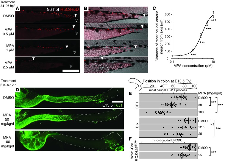
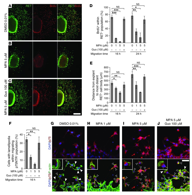
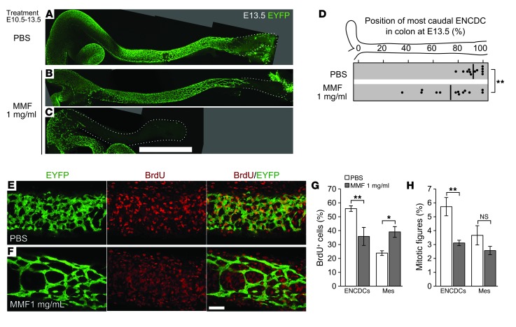
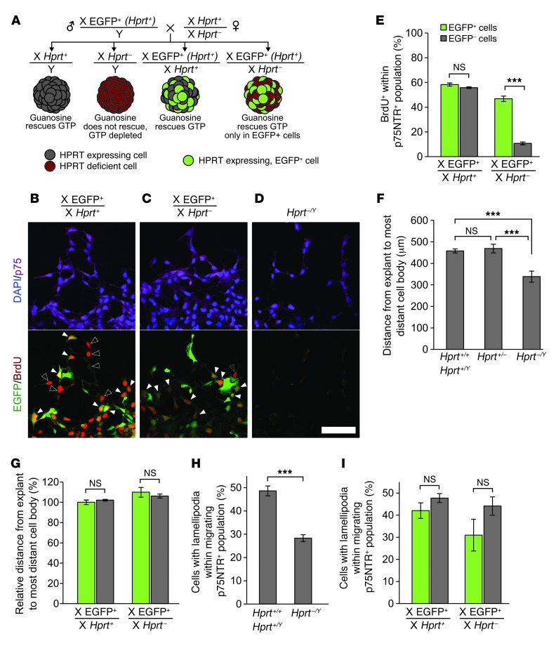
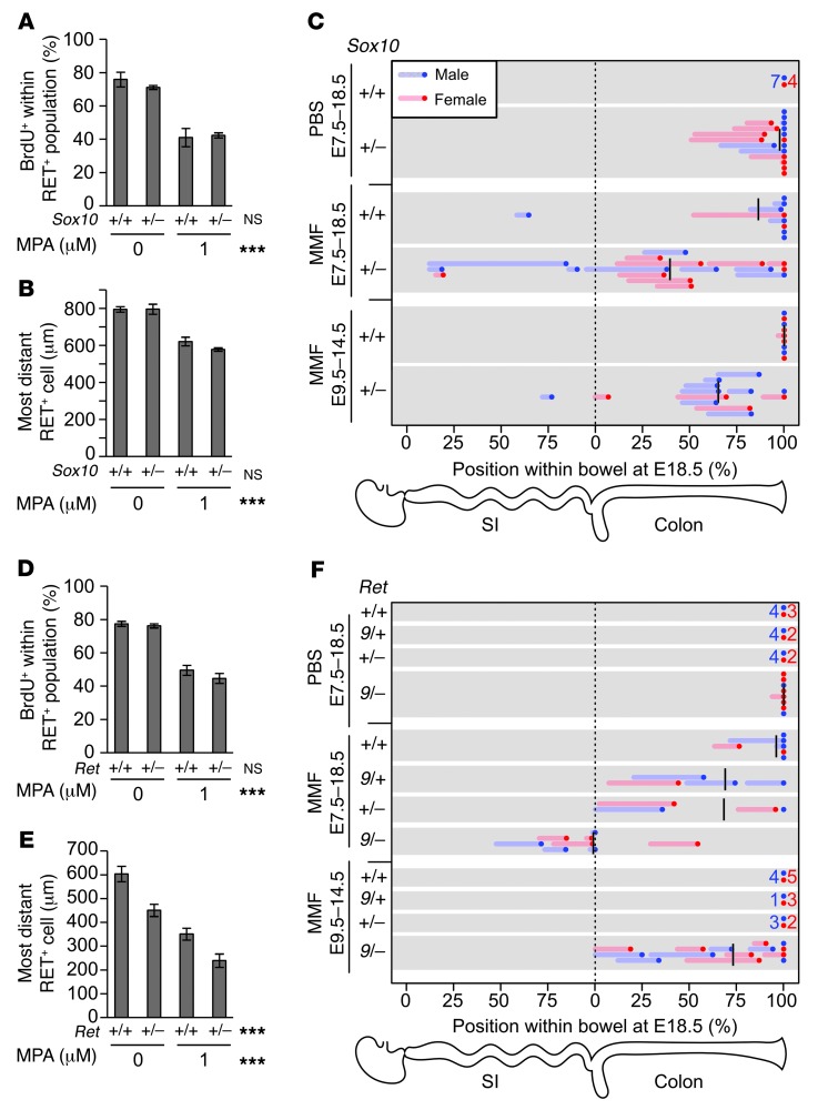
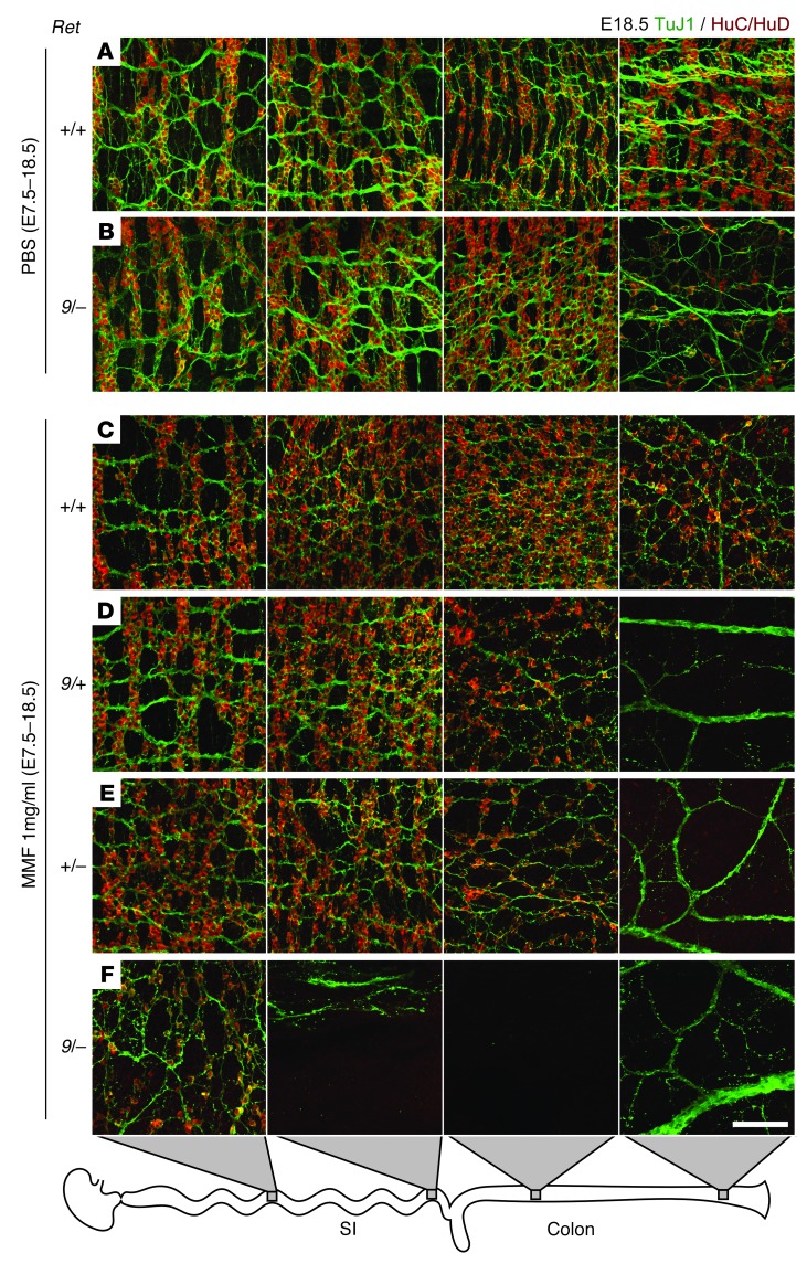
Hirschsprung disease (HSCR) is a partially penetrant oligogenic birth defect that occurs when enteric nervous system (ENS) precursors fail to colonize the distal bowel during early pregnancy. Genetic defects underlie HSCR, but much of the variability in the occurrence and severity of the birth defect remain unexplained. We hypothesized that nongenetic factors might contribute to disease development. Here we found that mycophenolate, an inhibitor of de novo guanine nucleotide biosynthesis, and 8 other drugs identified in a zebrafish screen impaired ENS development. In mice, mycophenolate treatment selectively impaired ENS precursor proliferation, delayed precursor migration, and induced bowel aganglionosis. In 2 different mouse models of HSCR, addition of mycophenolate increased the penetrance and severity of Hirschsprung-like pathology. Mycophenolate treatment also reduced ENS precursor migration as well as lamellipodia formation, proliferation, and survival in cultured enteric neural crest–derived cells. Using X-inactivation mosaicism for the purine salvage gene Hprt, we found that reduced ENS precursor proliferation most likely causes mycophenolate-induced migration defects and aganglionosis. To the best of our knowledge, mycophenolate is the first medicine identified that causes major ENS malformations and Hirschsprung-like pathology in a mammalian model. These studies demonstrate a critical role for de novo guanine nucleotide biosynthesis in ENS development and suggest that some cases of HSCR may be preventable.


Figures

Figure 1
Figure 1. MPA inhibited ENS development in developing zebrafish and mouse.
(AC) Developing WT zebrafish were exposed to DMSO or MPA from 34 to 96 hpf. (A) Larvae (N > 200) were immunostained for neuronal marker HuC/HuD. (B) Images in A merged with transmitted light. Filled arrowheads denote most caudal enteric neuron; open arrowheads denote vents. (C) Average uncolonized distal intestine, plotted vs. MPA dose and compared with control. (DF) MPA exposure by maternal intraperitoneal injection from E10.5 to E12.5 impaired enteric neuron colonization of the mouse hindgut at E13.5 (D), as visualized by the neuronal marker TuJ1 (left side, ileocecal junction; dotted line, colon outline). The position within each E13.5 colon of the most caudal (E) neuronal process (marked by TuJ1) or (F) ENCDC cell body (ascertained by the lineage marker EYFP or by SOX10 staining in EYFP littermates) in each E13.5 fetus is plotted for each MPA dose and mouse strain (thick lines denote mean). Scale bars: 250 μm (A and B); 1 mm (D). ***P < 0.001, Kolmogorov-Smirnov test (C); ANOVA and t test (E and F).
Figure 2
Figure 2. MPA reduced ENCDC migration, DNA synthesis, and lamellipodia in explant cultures.
(AC) Low-magnification confocal micrographs of 24-hour E12.5 midgut explant cultures immunostained for RET and BrdU (explant at left of each image). Guanosine (Guo; C) completely reversed the proliferation and migration reduction caused by MPA (B). (D) Quantification of BrdU labeling index and (E) distance migrated by the RET-expressing population after 16 and 24 hours in culture. (F) MPA reduced the percentage of cells with lamellipodia within the neural crest–derived cell population (stained with anti-p75NTR) most distant from the explant, an effect that was also reversed by guanosine. (GJ) Optical sections of p75NTR- and phalloidin-stained ENCDCs demonstrated the changes in cell shape associated with MPA treatment. Filled and open arrowheads denote ENCDCs with and without lamellipodium, respectively. Insets show details of ENCDCs at the leading edge (enlarged ×1.8). Scale bars: 250 μm (AC); 50 μm (GJ). *P < 0.05, **P < 0.01, ***P < 0.001, repeated-measures ANOVA (D and E); ANOVA (F).
Figure 3
Figure 3. MMF treatment reduces ENCDC migration and DNA synthesis in vivo.
(AD) Oral treatment of pregnant B6 dams with MMF from E10.5 to E13.5 reduced the colonization of the hindgut at E13.5. Shown are stitched maximum-intensity projections of untreated (A), mildly affected (B), and severely affected (C) fetal colons with EYFP-marked ENCDCs, demonstrating MMF’s inhibitory effect on ENCDC wavefront migration in vivo. Dotted lines denote outline of bowel. (D) The position within each E13.5 colon of the most caudal ENCDC cell body (ascertained by EYFP or by SOX10 staining in EYFP littermates) is plotted for each treatment (thick lines denote mean). (E and F) 8-μm-thick maximum-intensity projections of EYFP- and BrdU-labeled E13.5 colons. (G) Counting of BrdU+ cells within the volumes in F demonstrated a reduced proportion of BrdU+ ENCDCs and an increased proportion of BrdU+ mesenchymal cells after MMF treatment. mes, non-ENCDC mesenchyme. (H) Counting of mitotic figures showed that the proportion of ENCDCs undergoing mitosis was reduced, while the mitotic index of the mesenchyme was not significantly changed. Scale bars: 1 mm (AC); 50 μm (E and F). *P < 0.05, **P < 0.01, Student’s t test (D); ANOVA (G); ANOVA on log-transformed values (H).
Figure 4
Figure 4. Mosaic analysis reveals that effects of GTP depletion on migration and lamellipodia are non–cell autonomous.
(A) Mating scheme, genotypes, HPRT and EGFP expression patterns, and GTP depletion status of each population when cultured in the presence of both MPA and guanosine. These conditions create mixed cultures of GTP-depleted and EGFP-marked, guanosine-rescued ENCDCs in X-EGFP+, Hprt+/– explants, allowing migration of individual GTP-depleted ENCDCs to be examined in the context of a field of rescued ENCDCs. (BD) BrdU labeling revealed that guanosine rescued DNA synthesis in all ENCDCs in X-EGFP+, Hprt+/+ explants (B), but only rescued DNA synthesis within the HPRT-expressing ENCDCs marked by EGFP in X-EGFP+, Hprt+/– explants (C). As expected, guanosine failed to rescue DNA synthesis in Hprt–/Y ENCDCs (D). Filled arrowheads denote BrdU+EGFP+ double-positive ENCDCs; open arrowheads denote BrdU+GFP ENCDCs. (E) Quantification of BrdU labeling in mosaic explants. (F) ENCDC migration out of explants was impaired in Hprt–/Y explants, as expected, but Hprt+/+ and Hprt+/– explants produced similar ENCDC migration distances. (G) Quantification of migration within the depleted (EGFP) and rescued (EGFP+) populations of female Hprt+/– cells demonstrated that GTP-depleted cells did not migrate any less efficiently than rescued cells when surrounded by rescued cells. Similarly, while lamellipodia were reduced in Hprt–/Y explants (H), they were not reduced within the GTP-depleted ENCDC population in Hprt+/– explant cultures (I). Scale bar: 50 μm (BD). ***P < 0.001, paired t test (E and I); ANOVA (F); Wilcoxon signed-rank test (G); t test (H).
Figure 5
Figure 5. MMF treatment interacts with Ret and Sox10 mutations to increase penetrance and severity of HSCR-like pathology.
(A and D) MPA treatment in 24-hour explant cultures revealed that neither Ret nor Sox10 heterozygosity affected BrdU incorporation. (B and E) In contrast, Ret but not Sox10 heterozygosity reduced ENCDC migration distance, and MPA treatment had an additive effect on ENCDC migration distance. Interaction terms were not statistically significant. (C and F) Pregnant dams were provided MMF or control PBS in drinking water, and the ENS was examined at E18.5 with neuronal fiber (TuJ1) and soma (HuC/HuD) markers. The position of the most caudal soma within the intestine is plotted as a dot, and the region of hypoganglionosis is plotted as a line. Mean positions of aganglionosis are denoted by black lines. Groups without abnormal fetuses are summarized as 1 dot and number. Treatment with MMF from E7.5 to E18.5 resulted in hypoganglionosis and aganglionosis with genotype-dependant penetrance and severity. Treatment with MMF from E9.5 to E14.5 demonstrated genotype-dependent reversal of the MMF-induced developmental delays. ***P < 0.001, 2-way ANOVA (A, B, D, and E).
Figure 6
Figure 6. Representative maximum-intensity projections of myenteric plexus in the mid-small bowel, terminal ileum, proximal colon, and terminal colon from PBS-exposed WT (A) and Ret9/– (B) fetuses and MMF-treated Ret+/+ (C), Ret9/+ (D), Ret+/– (E), and Ret9/– (F) genotypes.
PBS-exposed Ret9/– colon (B) displayed distal hypoganglionosis. (DF) TuJ1 staining demonstrated thick nerve bundles in the aganglionic terminal colons and disorganized fibers in hypoganglionic regions. Scale bar: 100 μm (AF).

References

    1. Heuckeroth RO. Pediatric Neurogastroenterology. 2013. Hirschsprung disease. In: Faure C, Di Lorenzo C, Thapar N, eds. pp. 271–283. New York, New York, USA: Humana Press;
    1. Amiel J, et al. Hirschsprung disease, associated syndromes and genetics: a review. J Med Genet. 2008;45(1):1–14. doi: 10.1136/jmg.2007.053959. - DOI - PubMed
    1. Carrasquillo MM, McCallion AS, Puffenberger EG, Kashuk CS, Nouri N, Chakravarti A. Genome-wide association study and mouse model identify interaction between RET and EDNRB pathways in Hirschsprung disease. Nat Genet. 2002;32(2):237–244. doi: 10.1038/ng998. - DOI - PubMed
    1. Gabriel SB, et al. Segregation at three loci explains familial and population risk in Hirschsprung disease. Nat Genet. 2002;31(1):89–93. - PubMed
    1. Emison ES, et al. A common sex-dependent mutation in a RET enhancer underlies Hirschsprung disease risk. Nature. 2005;434(7035):857–863. doi: 10.1038/nature03467. - DOI - PubMed

Publication types

MeSH terms