. 2013 Nov;123(11):4769-80.
doi: 10.1172/JCI70528.

Chronic itch development in sensory neurons requires BRAF signaling pathways

Chronic itch development in sensory neurons requires BRAF signaling pathways

Zhong-Qiu Zhao et al. J Clin Invest. 2013 Nov.

Abstract

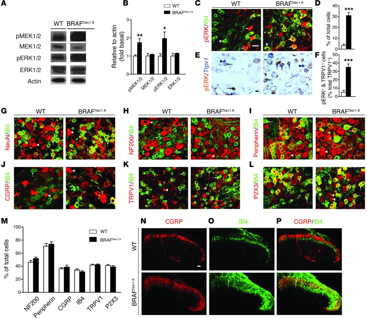
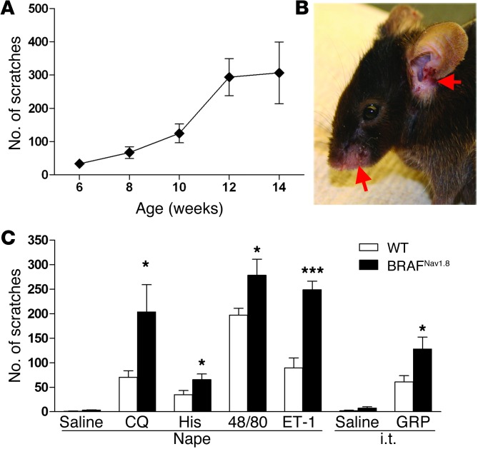
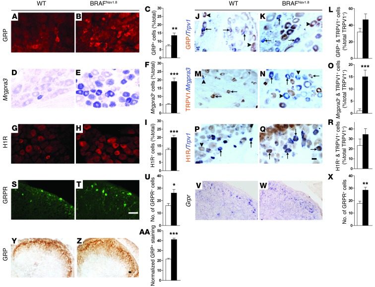
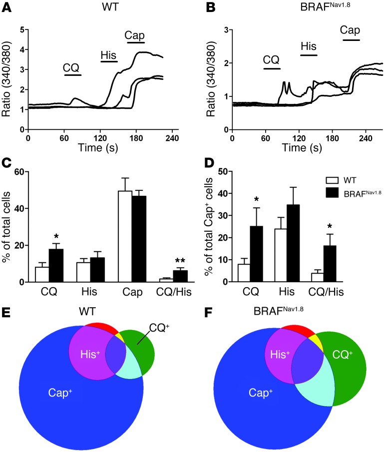
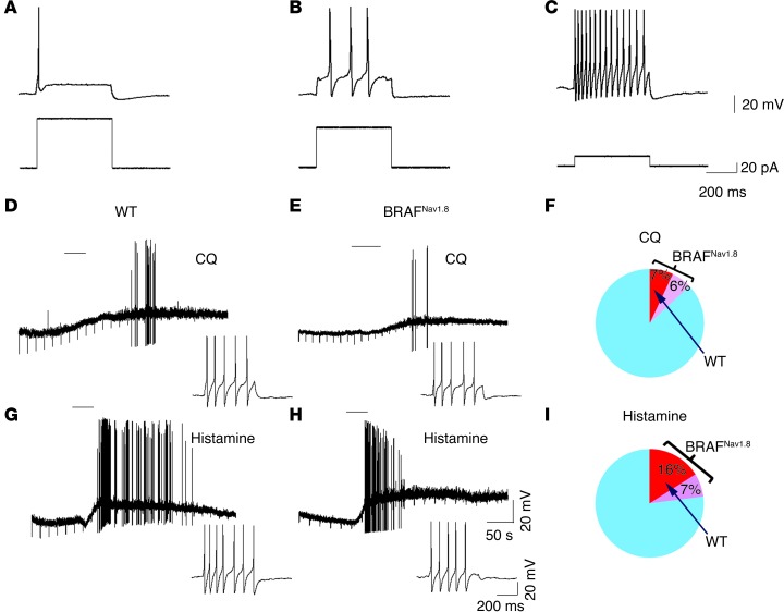
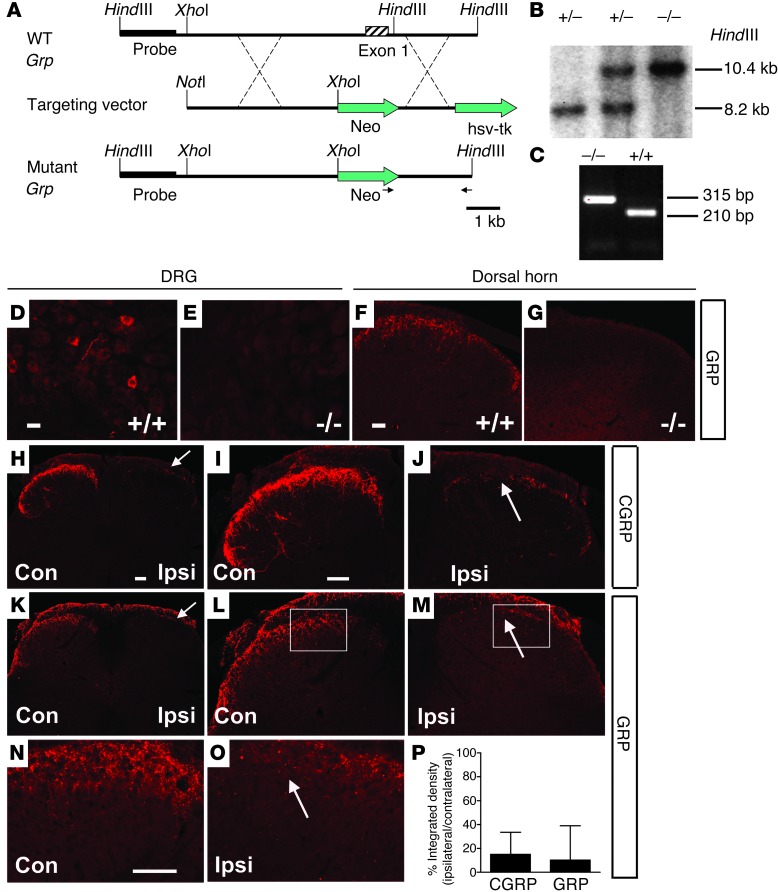
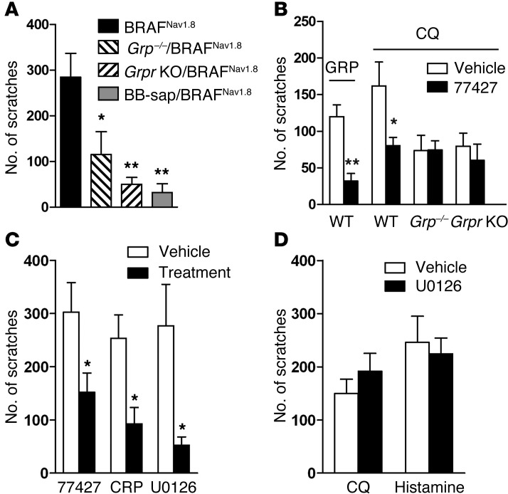
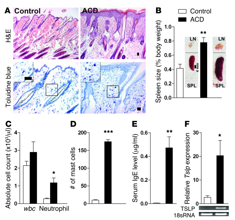
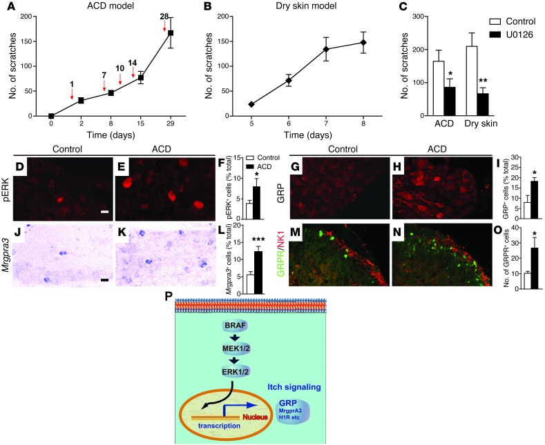
Chronic itch, or pruritus, is associated with a wide range of skin abnormalities. The mechanisms responsible for chronic itch induction and persistence remain unclear. We developed a mouse model in which a constitutively active form of the serine/threonine kinase BRAF was expressed in neurons gated by the sodium channel Nav1.8 (BRAF(Nav1.8) mice). We found that constitutive BRAF pathway activation in BRAF(Nav1.8) mice results in ectopic and enhanced expression of a cohort of itch-sensing genes, including gastrin-releasing peptide (GRP) and MAS-related GPCR member A3 (MRGPRA3), in nociceptors expressing transient receptor potential vanilloid 1 (TRPV1). BRAF(Nav1.8) mice showed de novo neuronal responsiveness to pruritogens, enhanced pruriceptor excitability, and heightened evoked and spontaneous scratching behavior. GRP receptor expression was increased in the spinal cord, indicating augmented coding capacity for itch subsequent to amplified pruriceptive inputs. Enhanced GRP expression and sustained ERK phosphorylation were observed in sensory neurons of mice with allergic contact dermatitis– or dry skin–elicited itch; however, spinal ERK activation was not required for maintaining central sensitization of itch. Inhibition of either BRAF or GRP signaling attenuated itch sensation in chronic itch mouse models. These data uncover RAF/MEK/ERK signaling as a key regulator that confers a subset of nociceptors with pruriceptive properties to initiate and maintain long-lasting itch sensation.


Figures

Figure 1
Figure 1. Increased levels of pMEK1/2 and pERK1/2 in sensory neurons and aberrant innervations in the spinal cords of BRAFNav1.8 mice.
(A) A representative Western blot and (B) quantified data show increased levels of pMEK1/2 and pERK1/2 but unchanged total MEK1/2 and ERK1/2 levels in DRGs of BRAFNav1.8 mice. n = 6 per genotype. All the lanes were run on the same gel but are noncontiguous. *P < 0.05, **P < 0.01, BRAFNav1.8 mice vs. wild-type. (CF) Increased pERK+ cells are indicated by double staining with pERK (red) and IB4 (green) or with pERK (brown) in Trpv1+ (blue) cells in BRAFNav1.8 mice compared to wild-type mice. n = 4 per genotype. ***P < 0.001. (GL) Expression of molecular markers with IB4 (green) in DRGs of wild-type and BRAFNav1.8 mice. (M) Quantitative comparison of DRG neurons between 2 groups. n = 3 per genotype. (NP) Expression of (N) CGRP and (O) IB4 in the spinal cords of wild-type and BRAFNav1.8 mice. Arrows and arrowheads indicate (C and E) pERK+ or pERK cells, respectively, and (GL) IB4+ or IB4 cells, respectively. Scale bar: 20 μm.
Figure 2
Figure 2. BRAFNav1.8 mice showed excessive spontaneous scratching and significantly enhanced pruritogen-evoked scratching.
(A) BRAFNav1.8 mice developed age-dependent spontaneous scratching behavior from 6 to 14 weeks. n = 6 to 8 per each time point. (B) BRAFNav1.8 mice developed skin lesions (arrows). (C) Prior to development of spontaneous scratching (at 4~6 weeks of age), BRAFNav1.8 mice showed significantly enhanced scratching responses to pruritogens injected into the nape (CQ; histamine [His]; compound 48/80 [48/80]; endothelin-1 [ET-1]) or to GRP injected i.t. Saline was the vehicle control. *P < 0.05, ***P < 0.001, BRAFNav1.8 compared to WT littermates in the same test, BRAFNav1.8 vs. wild-type. n = 5–9.
Figure 3
Figure 3. Ectopic expression of itch mediators in BRAFNav1.8 mice.
(AI) BRAFNav1.8 mice showed ectopic expression of (AC) GRP, (DF) Mrgpra3, and (GI) H1R in DRGs. (JR) BRAFNav1.8 mice showed increased expression of (MO) Mrgpra3 (blue) but not (JL) GRP (brown) and (PR) H1R (brown) in Trpv1+ cells (blue in JL and PR; brown in MO). (SX) Enhanced expression of GRPR as shown by (SU) GFP staining and (VX) Grpr mRNA in the dorsal horns of BRAFNav1.8 mice. (YAA) Enhanced GRP in primary afferents of BRAFNav1.8 mice. All mice were 8~12 weeks of age, except in VAA, where 2-week-old mice were used. Scale bar: 20 μm. (JQ) Arrows and arrowheads indicate TRPV1+ and TRPV1 neurons, respectively. *P < 0.05, **P < 0.01, ***P < 0.001, BRAFNav1.8 mice vs. wild-type. n = 4.
Figure 4
Figure 4. Enhanced and de novo responses to CQ and histamine but not to capsaicin in DRG cells from BRAFNav1.8 mice.
(A and B) Three representative traces of DRG cells from (A) wild-type or (B) BRAFNav1.8 mice responsive to CQ (100 μM), histamine (50 μM), and capsaicin (Cap; 1 μM). (B) Note that one CQ-responsive cell tested also responded to histamine and capsaicin. (C) The percentage of CQ-responsive DRG cells, but not histamine- and capsaicin-responsive cells, was significantly increased in BRAFNav1.8 mice. More DRG cells responded to both CQ and histamine in BRAFNav1.8 mice than in wild-type mice. (D) Of all capsaicin-sensitive cells in BRAFNav1.8 mice, there was a significant increase of cells that responded to CQ or to both CQ and histamine, whereas no difference was detected in the percentage of cells responsive to both capsaicin and histamine. (E and F) Venn diagrams showing the relative proportions of DRG cells of (E) wild-type mice and (F) BRAFNav1.8 mice responsive to CQ, histamine, and capsaicin. *P < 0.05, **P < 0.01, BRAFNav1.8 vs. wild-type. n = 4 (a total of 1,161 cells were tested).
Figure 5
Figure 5. Electrophysiological characterization of DRG cells from BRAFNav1.8 mice and wild-type mice.
(AC) The BRAFNav1.8 DRG cells exhibited altered distribution of firing properties. In both BRAFNav1.8 mutant and wild-type cells, 3 firing patterns were routinely observed as (A) SS, (B) delayed, and (C) tonic. (D, E, G, and H) Representative action potential firing patterns of wild-type and BRAFNav1.8 cells in response to (D and E) CQ and (G and H) histamine. The cells responding to pruritogens typically exhibited tonic firing properties with low rheobase values. Response magnitude was generally similar between wild-type and BRAFNav1.8 mice. (F and I) Venn diagrams of total DRG cells showing that the percentage changes of DRG cells responsive to (F) CQ and (I) histamine substantially increased in BRAFNav1.8 mice relative to wild-type mice. (F) CQ (1 mM) induced action potential firing in 3 out of 44 (6.8%) wild-type DRG cells and in 8 out of 61 (13.1%) mutant DRG cells. (I) Histamine (100 μM) induced action potential firing in 7 out of 44 (15.9%) wild-type DRG cells and in 14 out of 61 (23.0%) mutant DRG cells.
Figure 6
Figure 6. Generation of Grp–/– mice and confirmation of GRP expression in primary sensory neurons.
(A) Targeting strategy for generation of Grp–/– mice. (B and C) Germ line transmission was confirmed by (B) Southern blot and (C) PCR analysis. (DG) GRP expression in the (D and E) DRGs and (F and G) spinal cords of (D and F) wild-type mice and (E and G) Grp–/– mice. (HO) Expression of (HJ) CGRP and (KO) GRP in the lumbar spinal cords of C57BL/6J mice 14 days after unilateral dorsal rhizotomy (L5). On contralateral sides, both (H and I) CGRP+ and (K, L, and N) GRP+ fibers were mainly in the superficial dorsal horn (lamina I, IIo); but on the ipsilateral sides, both (H and J) CGRP and (K, M, and O) GRP staining was lost after unilateral L5 dorsal rhizotomy. Arrows indicate the elimination of staining. n = 3 per group. (P) Quantitation of remaining CGRP+ (15.1%) and GRP+ (10.2%) staining in the L5 superficial dorsal horn after rhizotomy. Boxed areas in L and M are shown at higher magnification in N and O. Scale bar: 10 μm (D and E); 20 μm (F and G); 40 μm (HO).
Figure 7
Figure 7. Genetic and pharmacologic blockade of spontaneous scratching behavior in BRAFNav1.8 mice.
(A) Spontaneous scratching of BRAFNav1.8 mice was significantly attenuated in Grp–/– or Grpr KO background and in mice treated with i.t. bombesin-saporin (BB-sap). *P < 0.05, **P < 0.01, 1-way ANOVA followed by post-hoc analysis, wild-type vs. BRAFNav1.8 mice. (B) GRP blocker 77427 (i.p.) significantly reduced scratching elicited by GRP (i.t.) or CQ (i.d.) in wild-type mice but not in Grp–/– or Grpr KO mice. (C) Spontaneous scratching of BRAFNav1.8 mice was significantly reduced by i.p. injection of 77427 or the H1R antagonist chlorpheniramine (CRP) or i.t. injection of a MEK inhibitor, U0126. (D) Scratching behavior of mice induced by i.d. CQ and histamine did not differ after i.t. U0126. *P < 0.05, **P < 0.01, 2-tailed t test vs. vehicle controls. n = 6–10.
Figure 8
Figure 8. Analysis of skin and immune system in adult mice with ACD.
(A) H&E and toluidine blue staining of the neck skin of the mice with ACD and the control mice. The mice with ACD showed epidermal hyperplasia, hyperkeratosis, acanthosis, dermal inflammation, and mast cell infiltration (insets show a few mast cells). Scale bar: 25 μm. (B and C) The systemic manifestations of allergic dermatitis were presented as highlighted by their (B) enlarged spleen and lymph nodes and (C) significantly increased blood neutrophil counts. SPL, spleen. Scale bar: 4 mm. (D) Quantitative analysis confirmed that ACD enhanced the extent of mast cell infiltration in the dermis. Mast cells in 9 random ×200 microscope fields were counted and averaged. (E) IgE levels were highly elevated in mice with ACD. (F) Tslp mRNA was overexpressed in the ACD skin. Representative RT-PCR images (bottom). *P < 0.05, **P < 0.01, ***P < 0.001, mice with ACD vs. the controls (acetone only). n = 6.
Figure 9
Figure 9. Mice with ACD- and dry skin–induced itch recapitulate the phenotype of BRAFNav1.8 mice.
(A) Mice with ACD induced by DNFB exhibited progressively increased scratching behavior. Red arrows indicate the time points when DNFB was applied, and spontaneous scratches were counted 24 hours afterward. n = 7. (B) Progressively increased scratching behavior was induced in mice with dry skin itch. n = 13. (C) Spontaneous scratching of mice with ACD or dry skin was significantly reduced by i.t. U0126. n = 5~9 per group. *P < 0.05, **P < 0.01, mice treated with U0126 vs. the controls. (DF) Ectopic activation of ERK and (GI) expression of GRP. (JL) Mrgpra3 in DRGs and (MO) GRPR as shown by GFP staining (E, H, K, and N) in the dorsal horns of mice with ACD compared to (D, G, J, and M) the controls (acetone only). In MO, GRPR-EGFP mice were used. Results in F, I, L, and O are quantitative comparisons between 2 groups (n = 3 per group). Scale bar: 10 μm (D, E, G, and H); 20 μm (J, K, M, and N). (P) Diagram illustrates BRAF/MEK/ERK regulation of itch molecular phenotype in DRG. *P < 0.05, ***P < 0.001, mice with ACD vs. the controls.

Similar articles

Cited by

References

    1. Imamachi N, et al. TRPV1-expressing primary afferents generate behavioral responses to pruritogens via multiple mechanisms. Proc Natl Acad Sci U S A. 2009;106(27):11330–11335. doi: 10.1073/pnas.0905605106. - DOI - PMC - PubMed
    1. Andrew D, Craig AD. Spinothalamic lamina I neurons selectively sensitive to histamine: a central neural pathway for itch. Nat Neurosci. 2001;4(1):72–77. - PubMed
    1. Davidson S, Giesler GJ. The multiple pathways for itch and their interactions with pain. Trends Neurosci. 2010;33(12):550–558. doi: 10.1016/j.tins.2010.09.002. - DOI - PMC - PubMed
    1. Ross SE. Pain and itch: insights into the neural circuits of aversive somatosensation in health and disease. Curr Opin Neurobiol. 2011;21(6):880–887. doi: 10.1016/j.conb.2011.10.012. - DOI - PubMed
    1. Jeffry J, Kim S, Chen ZF. Itch signaling in the nervous system. Physiology (Bethesda). 2011;26(4):286–292. doi: 10.1152/physiol.00007.2011. - DOI - PMC - PubMed

Publication types

MeSH terms