

doi: 10.1038/leu.2013.298.
Epub 2013 Oct 18.
The tumor suppressor axis p53/miR-34a regulates Axl expression in B-cell chronic lymphocytic leukemia: implications for therapy in p53-defective CLL patients
Affiliations
- PMID: 24217154
- PMCID: PMC3929965
- DOI: 10.1038/leu.2013.298
Item in Clipboard
The tumor suppressor axis p53/miR-34a regulates Axl expression in B-cell chronic lymphocytic leukemia: implications for therapy in p53-defective CLL patients
Leukemia.
2014 Feb.
No abstract available
Conflict of interest statement
Authors declare no potential conflict of interest
Figures
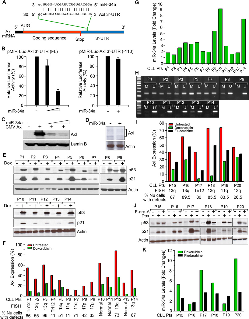
A. Alignment of Axl 3’-UTR with miR-34a sequence. B. miR-34a targets Axl 3’-UTR. HEK293 cells were co-transfected with a luciferase reporter plasmid DNAs containing the entire Axl 3’-UTR with a putative miR-34a-binding site (full-length) or without the miR-34a-binding site [Axl 3’-UTR (-110)] and increasing amounts of miR-34a mimic or sc- miR as control. After 24 hour, luciferase activity was measured in the cell lysates. Experiments were repeated twice in triplicates. Data were normalized and presented as mean values with standard deviations. C. Exogenous miR-34a reduces Axl expression. HEK293 cells were co-transfected with an Axl expressing plasmid DNA containing the putative miR-34a binding site in the 3’-UTR and increasing amounts of miR-34a or sc-miR for 24 hours. Cells were examined for the expression of Axl by Western blot using a specific antibody to Axl. Lamin B was used as a loading control. D. Introduction of miR-34a mimic reduces Axl expression in primary CLL B-cells. Purified CLL B-cells were transfected with miR-34a mimic or sc-miR as control using B-cell specific nucleofection reagent (Amaxa). After 24 hours, cell lysates were analyzed for the expression of Axl by Western blots using a specific antibody. Actin was used as a loading control. Representative result of CLL B-cells from 3 different CLL patients is shown. E. Doxorubicin activates p53. Lysates from purified CLL B-cells treated with doxorubicin or left untreated for 16 hours were analyzed for the expression of p53 and its downstream target p21 in Western blots using specific antibodies. Actin was used as a loading control. CLL B-cells from various CLL patients are indicated by arbitrary numbers (P1-P14). F. Impact of doxorubicin on Axl expression. CLL B-cells from the same CLL patients’ cohort (P1-P14) studied above (panel E) were treated with doxorubicin or left untreated for 16 hours. Axl expression was determined by flow cytometry using a specific antibody. Percent of cells with FISH detectable chromosomal abnormalities are indicated. G. Doxorubicin treatment activates miR-34a expression in CLL B-cells. Total RNA was extracted from untreated and doxorubicin-treated CLL B-cells and miR-34a levels were measured by qRT-PCR using primers specific for mature miR-34a and normalized using the 2-∆∆Ct method relative to U6-snRNA (RNU6B). Relative miR-34a expression levels in untreated vs. doxorubicin-treated CLL B-cells are presented as “fold changes” determined as mean of triplicate values. CLL B-cells studied here were chosen from the same cohort of CLL patients depicted and studied above in panels E&F. H. Methylation specific-PCR analysis of the miR-34a promoter in CLL B-cells. DNA isolated from purified CLL B-cells of the CLL patients (P1 - P3, P5, P7 - P14) whose miR-34a levels were studied above (panel G) were subjected to bisulfite conversion and analyzed by methylation specific-PCR (MSP) with primers specific for methylated (M) and unmethylated (U) miR-34a promoter DNA. Amplified PCR products were run on a 1% agarose gel along with a standard DNA ladder (Invitrogen; 1st lane from left) to ascertain the size of the PCR products. I. In vitro Fludarabine treatment reduces Axl expression on CLL B-cells. CLL B-cells from CLL patients (P15 – P20) were treated with fludarabine (F-ara-A) for 16 hour or left untreated and Axl expression was determined by flow cytometry as described above. Doxorubicin was included as a positive control. J. Fludarabine activates p53. Cell lysates prepared from untreated, doxorubicin-treated and F-ara-A-treated CLL B-cells from CLL patients (P15 – P20) were examined for the expression of p53 and p21 in Western blot analysis. Actin was used as a loading control. K. Fludarabine upregulates miR-34a expression. Total RNA was extracted from untreated, doxorubicin-treated and F-ara-A-treated CLL B-cells from the same CLL patients used above (P15 – P20). Mature miR-34a levels were measured by qRT-PCR and presented as “fold changes” as described in panel G.
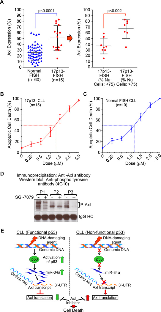
A. CLL B-cells with 17p13-defects express significantly higher levels of Axl. CLL B-cells from patients with 17p13-chromosomal defects (n=15) or no FISH detectable chromosomal abnormalities (n=60) were examined for Axl expression by flow cytometry using a specific antibody. Results demonstrate significantly increased levels (p<0.0001) of Axl on CLL B-cells with 17p13-defects as compared to that on CLL B-cells with no FISH detectable chromosomal defects (normal FISH). Further analysis demonstrates that the CLL patients containing >75% 17p13-defective CLL clones express significantly higher levels (<0.002) of Axl as compared to those having <75% 17p13-defective leukemic B-cells (right panel). B & C.
Axl inhibition induces robust apoptosis in CLL B-cells. CLL B-cells from CLL patients with 17p13 chromosomal abnormalities (panel B) or normal FISH (panel C) were treated with increasing doses of the high-affinity Axl-inhibitor, SGI-7079 for 72 hours. Cells were harvested and induction of apoptosis was determined by flow cytometric analysis after staining with Annexin FITC and propidium iodide. Results are presented as mean values with standard deviations. D. SGI-7079 targets Axl phosphorylation in CLL B-cells. Purified CLL B-cells from CLL patients were treated with a sub-lethal dose of SGI-7079 for 16 hours. Axl was immunoprecipitated from equal amount of cell lysates and phosphorylation status was examined by Western blot analysis using a phospho-tyrosine-specific antibody (4G10-Platinum). Vehicle-treated cells were used as controls. IgG heavy chain is shown as a loading control. Results show a substantial level of reduction in phosphorylation of Axl in CLL B-cells following treatment with SGI-7079. CLL patients are indicated by arbitrary numbers (P1 – P3). E. Model of p53-mediated regulation of Axl in CLL B-cells. Activation of p53 via various stressors in CLL with a functional p53 inhibits translation of the Axl mRNA by activating miR-34a transcription. Accumulated miR-34a binds the single binding site on the Axl 3’-UTR and inhibits its translation resulting in reduction of Axl protein level. In CLL B-cells with a non-functional p53 (due to chromosomal deletion and/or inactivating mutation on p53 gene) this regulatory pathway remains non-functional which likely results in significant upregulation/stabilization of Axl RTK protein levels. Importantly, this study finds that Axl has the potential to be an effective therapeutic target in CLL B-cells with or without a functional p53 gene.
References
-
- Ghosh AK, Secreto C, Boysen J, Sassoon T, Shanafelt TD, Mukhopadhyay D, et al. The novel receptor tyrosine kinase Axl is constitutively active in B-cell chronic lymphocytic leukemia and acts as a docking site of nonreceptor kinases: implications for therapy. Blood. 2011 Feb 10;117(6):1928–1937. - PMC - PubMed
-
- Hermeking H. The miR-34 family in cancer and apoptosis. Cell Death Differ. 2010 Feb;17(2):193–199. - PubMed
-
- Ghosh AK, Boysen J, Price-troska T, Secreto C, Zent CS, Kay N. Axl Receptor Tyrosine Kinase Signaling Pathway and the p53 Tumor Suppressor Protein Exist In A Novel Regulatory Loop In B-Cell Chronic Lymphocytic Leukemia Cells. ASH Annual Meeting Abstracts. 2011 Nov 18;118(21):799. 2011.
-
- Mudduluru G, Ceppi P, Kumarswamy R, Scagliotti GV, Papotti M, Allgayer H. Regulation of Axl receptor tyrosine kinase expression by miR-34a and miR-199a/b in solid cancer. Oncogene. 2011 Jun 23;30(25):2888–2899. - PubMed
Publication types
MeSH terms
Substances
Grants and funding
LinkOut - more resources
Full Text Sources
Other Literature Sources
Research Materials
Miscellaneous
