

Investigation of a method for generating synthetic CT models from MRI scans of the head and neck for radiation therapy
- PMID: 24217183
- PMCID: PMC3886820
- DOI: 10.1088/0031-9155/58/23/8419
Investigation of a method for generating synthetic CT models from MRI scans of the head and neck for radiation therapy
Abstract
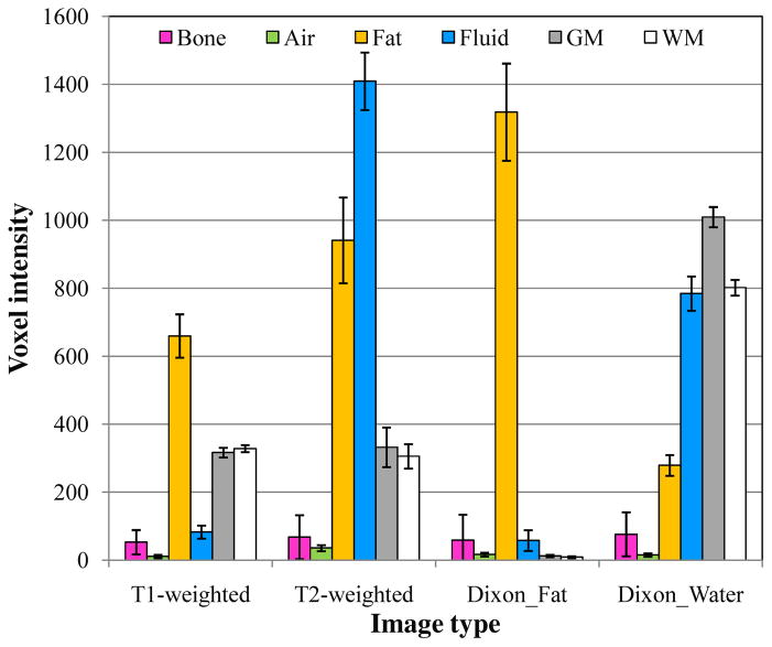
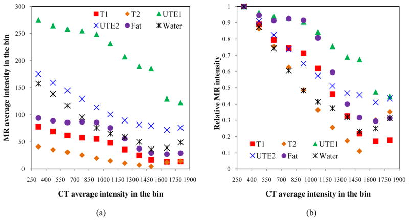
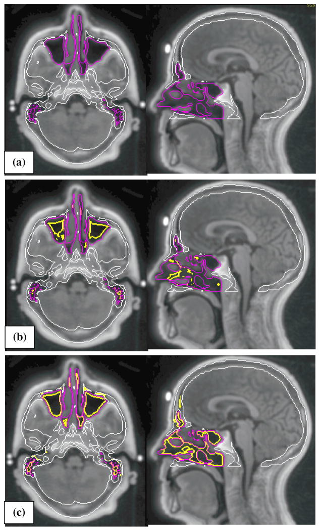
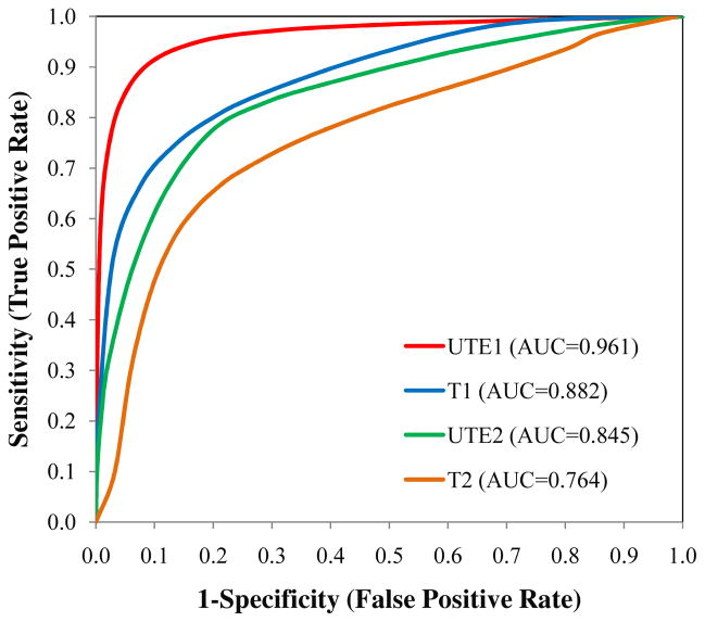
Magnetic resonance (MR) images often provide superior anatomic and functional information over computed tomography (CT) images, but generally are not used alone without CT images for radiotherapy treatment planning and image guidance. This study aims to investigate the potential of probabilistic classification of voxels from multiple MRI contrasts to generate synthetic CT ('MRCT') images. The method consists of (1) acquiring multiple MRI volumes: T1-weighted, T2-weighted, two echoes from a ultra-short echo time (UTE) sequence, and calculated fat and water image volumes using a Dixon method, (2) classifying tissues using fuzzy c-means clustering with a spatial constraint, (3) assigning attenuation properties with weights based on the probability of individual tissue classes being present in each voxel, and (4) generating a MRCT image volume from the sum of attenuation properties in each voxel. The capability of each MRI contrast to differentiate tissues of interest was investigated based on a retrospective analysis of ten patients. For one prospective patient, the correlation of skull intensities between CT and MR was investigated, the discriminatory power of MRI in separating air from bone was evaluated, and the generated MRCT image volume was qualitatively evaluated. Our analyses showed that one MRI volume was not sufficient to separate all tissue types, and T2-weighted images was more sensitive to bone density variation compared to other MRI image types. The short echo UTE image showed significant improvement in contrasting air versus bone, but could not completely separate air from bone without false labeling. Generated MRCT and CT images showed similar contrast between bone and soft/solid tissues. These results demonstrate the potential of the presented method to generate synthetic CT images to support the workflow of radiation oncology treatment planning and image guidance.
Figures


References
-
- Barra V, Boire JY. Tissue segmentation on MR images of the brain by possibilistic clustering on a 3D wavelet representation. J Magn Reson Imaging. 2000;11:267–78. - PubMed
-
- Berker Y, Franke J, Salomon A, Palmowski M, Donker HC, Temur Y, Mottaghy FM, Kuhl C, Izquierdo-Garcia D, Fayad ZA, Kiessling F, Schulz V. MRI-based attenuation correction for hybrid PET/MRI systems: a 4-class tissue segmentation technique using a combined ultrashort-echo-time/Dixon MRI sequence. J Nucl Med. 2012;53:796–804. - PubMed
-
- Chen L, Nguyen TB, Jones E, Chen Z, Luo W, Wang L, Price RA, Jr, Pollack A, Ma CM. Magnetic resonance-based treatment planning for prostate intensity-modulated radiotherapy: creation of digitally reconstructed radiographs. Int J Radiat Oncol Biol Phys. 2007;68:903–11. - PubMed
-
- Chen S, Zhang D. Robust image segmentation using FCM with spatial constraints based on new kernel-induced distance measure. IEEE Trans Syst Man Cybern B Cybern. 2004;34:1907–16. - PubMed
Publication types
MeSH terms
Grants and funding
LinkOut - more resources
Full Text Sources
Other Literature Sources
Medical