. 2013 Nov 8;5(11):2093-108.
doi: 10.3390/toxins5112093.

Oral toxicity of okadaic acid in mice: study of lethality, organ damage, distribution and effects on detoxifying gene expression

Affiliations

Oral toxicity of okadaic acid in mice: study of lethality, organ damage, distribution and effects on detoxifying gene expression

Andres C Vieira et al. Toxins (Basel). .

Abstract

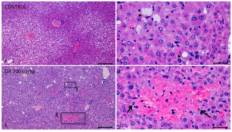
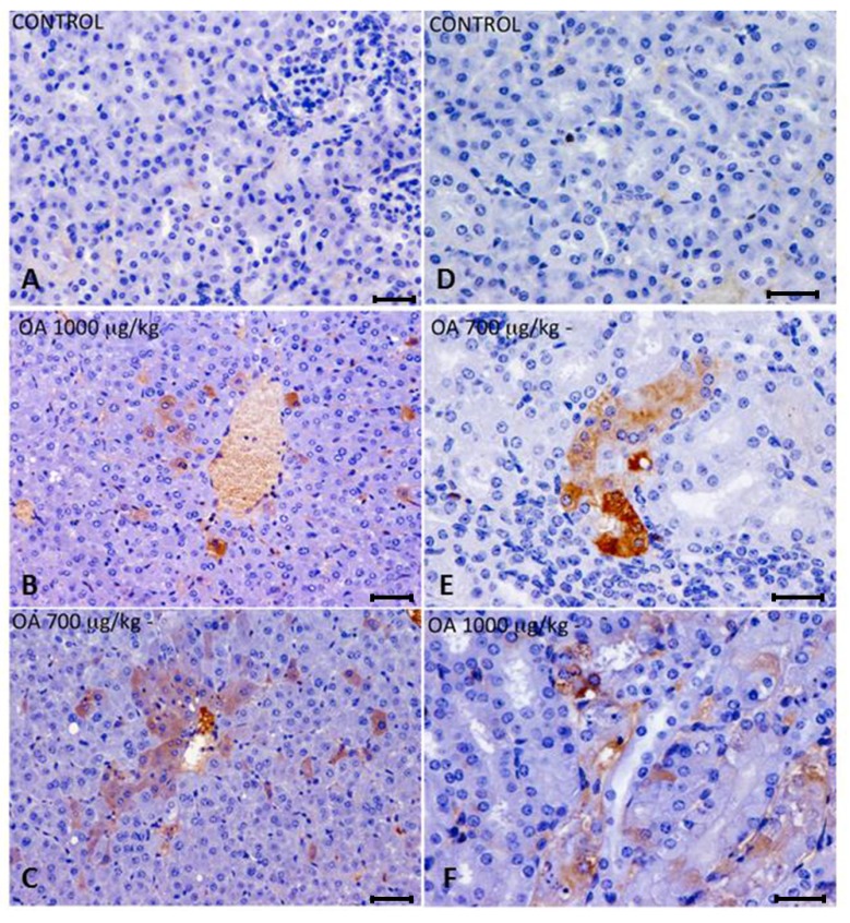
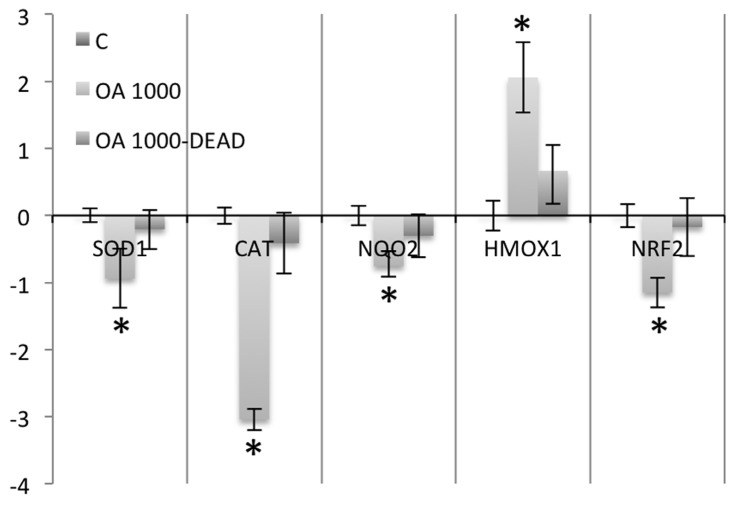
In vivo, after administration by gavage to mice and rats, okadaic acid has been reported to produce lesions in liver, small intestine and forestomach. Because several reports differ in the damage detected in different organs, and on okadaic acid distribution after consumption, we determined the toxicity of this compound after oral administration to mice. After 24 hours, histopathological examination showed necrotic foci and lipid vacuoles in the livers of intoxicated animals. By immunohistochemical analysis, we detected this toxin in the liver and kidneys of intoxicated animals. Okadaic acid induces oxidative stress and can be activated in vitro into reactive compounds by the post-mitochondrial S9 fraction, so we studied the okadaic effect on the gene expression of antioxidant and phase II detoxifying enzymes in liver. We observed a downregulation in the expression of these enzymes and a reduction of protein expression of catalase and superoxide dismutase 1 in intoxicated animals.


Figures

Figure 1
Figure 1
Photomicrographs of liver sections from both control and treated animals. (AD) Photomicrographs of liver sections from control (A) and 700 μg/kg OA-intoxicated mice (BD) showing necrotic areas in liver of treated animals (B). (C) Amplified area of photomicrograph B (1). Neighboring hepatocytes to the necrotic lesions showed cellular swelling, lipid vacuoles of different sizes (arrow heads) and either pleomorphic or pyknotic nuclei. (D) Amplified area of photomicrograph B (2). Multifocal aggregates of necrotic hepatocytes randomly located with the consequent dilation of the sinusoids, and polymorphonuclear inflammatory infiltrates (arrows) could be observed. Scale bars, 200 μm (A and B) and 20 μm (C and D).
Figure 2
Figure 2
(AC) Immunohistochemistry of hepatic sections of control and OA-intoxicated mice. (A) Control liver section. (B,C) OA detected was distributed throughout the liver parenchyma and also concentrated around centrilobular areas in 1000 µg/kg (B) and 700 µg/kg (C) treated specimens. (DF) Immunohistochemistry of kidney sections of control and OA-intoxicated mice. (D) Control kidney section.(E) 700 µg/kg treated specimens, showing strong immunoreactivity in proximal convoluted tubules. (F) 1000 µ/kg treated mice showing that immunoreactivity against OA was distributed in a scattered manner in proximal tubules. Cytoplasm of tubular epithelial cells was vacuolated and nuclear changes were evident.Scale bars 60 μm (AC) and 20 μm (DF).
Figure 3
Figure 3
Real-time polymerase chain reaction (PCR) of liver mRNA, from control and OA-treated mice. (A) A decrease in the expression of SOD1 and CAT was observed. While the levels of NQO2 mRNA fell in livers of OA-treated animals, an increase in the expression of HMOX2 and GPX2 was observed. There was also a decrease in the expression of the transcription factor Nrf2 in OA-intoxicated animals. (B) Western blots of CAT, SOD1 and β tubulin (loading control) of control (−) and OA-treated mice (+). (C) Densitometry analysis of the Western blot results showing downregulation of CAT and SOD1 in animals intoxicated with 700 µg/kg OA. * Significant differences with respective controls (p < 0.05, n = 2).
Figure 4
Figure 4
Real-time PCR comparing SOD1, CAT, NQO2, HMOX1 and NRF2 expression in nine mice that survived 1000 μg/kg OA-intoxication (24 hours) with three animals that died early after intoxication (between the hours 3 and 5) with the same dose of toxin.
Figure 5
Figure 5
Scheme describing the design of the OA-intoxication experiment and the times when samples for analysis were collected. Scheme A describes an experiment repeated three times, while scheme B describes a unique experiment.

References

    1. Yasumoto T., Murata M., Oshima Y., Sano M., Matsumoto G., Clardy J. Diarrhetic shellfish toxins. Tetrahedron. 1985;41:1019–1025. doi: 10.1016/S0040-4020(01)96469-5. - DOI
    1. Vale P., Sampayo M.A. Domoic acid in Portuguese shellfish and fish. Toxicon. 2001;39:893–904. doi: 10.1016/S0041-0101(00)00229-4. - DOI - PubMed
    1. Yasumoto T., Oshima Y., Yamaguchi M. Occurrence of a new type of shellfish poisoning in the Tohoku district. Bull. Jpn. Soc. Sci. Fish. 1978;46:1249–1275. doi: 10.2331/suisan.44.1249. - DOI
    1. Fujiki H., Suganuma M. Unique features of the okadaic acid activity class of tumor promoters. J. Cancer Res. Clin. Oncol. 1999;125:150–155. doi: 10.1007/s004320050257. - DOI - PubMed
    1. Bialojan C., Takai A. Inhibitory effect of a marine-sponge toxin, okadaic acid, on protein phosphatases. Specificity and kinetics. Biochem. J. 1988;256:283–290. - PMC - PubMed

Publication types

MeSH terms

LinkOut - more resources