. 2014 Feb;42(3):1606-18.
doi: 10.1093/nar/gkt1096. Epub 2013 Nov 11.

Cellular senescence mediated by p16INK4A-coupled miRNA pathways

Affiliations

Cellular senescence mediated by p16INK4A-coupled miRNA pathways

Marita G Overhoff et al. Nucleic Acids Res. 2014 Feb.

Abstract

p16 is a key regulator of cellular senescence, yet the drivers of this stable state of proliferative arrest are not well understood. Here, we identify 22 senescence-associated microRNAs (SA-miRNAs) in normal human mammary epithelial cells. We show that SA-miRNAs-26b, 181a, 210 and 424 function in concert to directly repress expression of Polycomb group (PcG) proteins CBX7, embryonic ectoderm development (EED), enhancer of zeste homologue 2 (EZH2) and suppressor of zeste 12 homologue (Suz12), thereby activating p16. We demonstrate the existence of a tight positive feedback loop in which SA-miRNAs activate and re-enforce the expression of other SA-miRNA members. In contrast, PcG members restrain senescence by epigenetically repressing the expression of these SA-miRNAs. Importantly, loss of p16 leads to repression of SA-miRNA expression, intimately coupling this effector of senescence to the SA-miRNA/PcG self-regulatory loop. Taken together, our findings illuminate an important regulatory axis that underpins the transition from proliferation to cellular senescence.


Figures
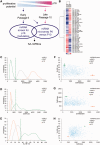
Figure 1.
Figure 1.
miRNA library screen and miRNA expression profiling identify SA-miRNAs. (A) Schematic representation of the strategy to identify SA-miRNAs. (B) Heatmap diagram of the results of two-way hierarchical clustering of miRNAs for HMEC P6 and HMEC P10. The clustering was performed on log2 (Hy3/Hy5) rations, which passed the filtering criteria on variation across samples. The relative expression level of each miRNA across all samples is shown: red represents an expression level above the mean and blue expression lower than the mean. (C–E) Frequency distributions of (C) cell number, (D) cell area and (E) percentage of p16 positive cells (% p16 +ve) following transfection with siGLO (grey), CBX7 (green) or p16 siRNA (orange) obtained at the time of the miRNA screen for modulators of p16-mediated cellular senescence. Cut-off values for each measure that were used to classify SA-miRNAs (green dotted lines). (F–H) Scatter plots illustrating phenotypic criteria data for each control siRNA as per (C–E) and the miRNA screening data (blue). (F) Cell number versus percentage of p16 positive cells (% p16 +ve), (G) cell number versus cell area and (H) cell area versus percentage p16 positive cells.
Figure 2.
Figure 2.
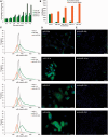
SA-miRNAs function during, and promote, cellular senescence by inducing p16. (A) SA-miRNA expression during cellular senescence in normal finite-lifespan HMECs from P6 to P12. (B) Cellular proliferation 5 days following transfection of HMEC P6 with the indicated SA-miRNA (60 nM; green) or anti-SA-miRNA (90 nM; orange). (C) Frequency distribution of p16 intensity following transfection with siGLO negative control (grey), the corresponding SA-miRNA (green) or anti-SA-miRNA (orange). (D, E) HMECs stained with 4′,6-diamidino-2-phenylindole (blue) and αp16 (green) following transfection with (D) SA-miRNA or (E) anti-SA-miRNA. Assays were performed in triplicates, and means ± SD from three independent experiments are shown.
Figure 3.
Figure 3.
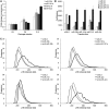
SA-miRNAs function during, and promote, cellular senescence by inducing p16 in fibroblasts. (A) SA-miRNA expression during cellular senescence in human fibroblasts from P6 to P12. (B) Cellular proliferation following transfection of fibroblasts P6 with the indicated SA-miRNA (60 nM; white, light grey) or anti-SA-miRNA (90 nM; dark grey, black) at day 2 (D2) and day 5 (D5) post transfection. (C) Frequency distribution of p16 intensity 5 days following transfection with siGLO negative control (grey), the corresponding SA-miRNA (solid black) or anti-SA-miRNA (dashed black). Assays were performed in triplicates, and means ± SD from three independent experiments are shown.
Figure 4.
Figure 4.
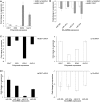
SA-miRNAs directly and coordinately regulate PcG genes. (A) miRSVR score analysis predicts potential binding sites for SA-miRNAs in the 3′UTR of multiple PcG mRNAs (see Supplementary Figure S1). Luciferase reporter assays with wild-type 3′ UTR constructs of CBX7, EED, EZH2 or Suz12 demonstrate that SA-miR-26b, 181a, 210 and 424 repress PcG protein activity. (B) Luciferase reporter assays with mutated 3′UTR constructs as described in Supplementary Table S1. Values for mock transfections were normalized to 1. (C) PcG protein expression declines during cellular senescence. (D, E) qRT-PCR quantitation of PcG expression following transfection of HMEC P6 with the indicated (D) SA-miRNA or (E) anti-SA-miRNA. Assays were performed in triplicates, and means ± SD from three independent experiments are shown.
Figure 5.
Figure 5.
p16, PcGs and the SA-miRNAs are interconnected in coupled feed-back loops. (A and B) Fold change in SA-miRNA expression following the transfection of HMEC P6 with the indicated (A) SA-miRNA or (B) anti-SA-miRNA. (C and D) ChIP analysis of H3K27Me3 at (C) the indicated regions of the INK/ARF locus, and (D) the genomic loci of SA-miRNAs following stable overexpression of CBX7 (HMEC.CBX7) in HMEC P6 and HMEC P11 cells. ChIP analysis of GAPDH and miR-876 serves as negative controls. A map to show where the primers align is given for INK/ARF locus in the top (C) and for the SA-miRNAs in Supplementary Table S4. Assays were performed in triplicates, and means ± SD from three independent experiments are shown.
Figure 6.
Figure 6.
p16 coupled to SA-miRNA and PcG regulatory mechanism. (A and B) Fold change in (A) PcG and (B) SA-miRNA expression following stable knockdown of p16 expression (HMEC.p16shRNA) or stable CBX7 overexpression (HMEC.CBX7). Data are normalized to HMEC.Vector control. NS = not shown. The fold change in CBX7 expression in the HMEC.CBX7 relative to HMEC.Vector was +1294 ± 58.96. (C and D) Fold change in (C) Polycomb members and (D) SA-miRNA expression following transient knockdown of CBX7 (green, left) or p16 expression (orange, right). Data expressed relative to the siGLO negative control. Assays were performed in triplicates, and means ± SD from three independent experiments are shown.
Figure 7.
Figure 7.
p16-coupled miRNA pathways mediate cellular senescence. PcG members CBX7, EED, EZH2 and Suz12 function to restrain cellular senescence by epigenetically repressing the expression of SA-miRNAs, SA-miRNA-26b, 181a, 210 and 424, as well as p16. CBX7 functions to positively promote the expression of other PcG members. The onset of cellular senescence disrupts this equilibrium, and SA-miRNA expression is stimulated. SA-miRNAs directly target PcG mRNAs for degradation. This, together with SA-miRNA cross-talk, ensures continued expression of the SA-miRNA signature, and as a direct consequence, PcG-mediated repression of p16 is relieved and the senescence programme enforced. p16, in turn, ensures self-reinforcement through a positive feedback loop with SA-miRNAs and a negative feedback loop with PcG members.

References

    1. Hayflick L. The limited in vitro lifetime of human diploid cell strains. Exp. Cell Res. 1965;37:614–636. - PubMed
    1. Michaloglou C, Vredeveld LC, Soengas MS, Denoyelle C, Kuilman T, van der Horst CM, Majoor DM, Shay JW, Mooi WJ, Peeper DS. BRAFE600-associated senescence-like cell cycle arrest of human naevi. Nature. 2005;436:720–724. - PubMed
    1. Krishnamurthy J, Torrice C, Ramsey MR, Kovalev GI, Al-Regaiey K, Su L, Sharpless NE. Ink4a/Arf expression is a biomarker of aging. J. Clin. Invest. 2004;114:1299–1307. - PMC - PubMed
    1. Campisi J, d'Adda di Fagagna F. Cellular senescence: when bad things happen to good cells. Nat. Rev. Mol. Cell Biol. 2007;8:729–740. - PubMed
    1. Scott LJ, Mohlke KL, Bonnycastle LL, Willer CJ, Li Y, Duren WL, Erdos MR, Stringham HM, Chines PS, Jackson AU, et al. A genome-wide association study of type 2 diabetes in Finns detects multiple susceptibility variants. Science. 2007;316:1341–1345. - PMC - PubMed

Publication types

Substances