Review
. 2014 Jan-Feb;5(1):111-30.
doi: 10.1002/wrna.1201. Epub 2013 Nov 11.

Finding the target sites of RNA-binding proteins

Affiliations
Free PMC article
Review

Finding the target sites of RNA-binding proteins

Xiao Li et al. Wiley Interdiscip Rev RNA. 2014 Jan-Feb.
Free PMC article

Abstract

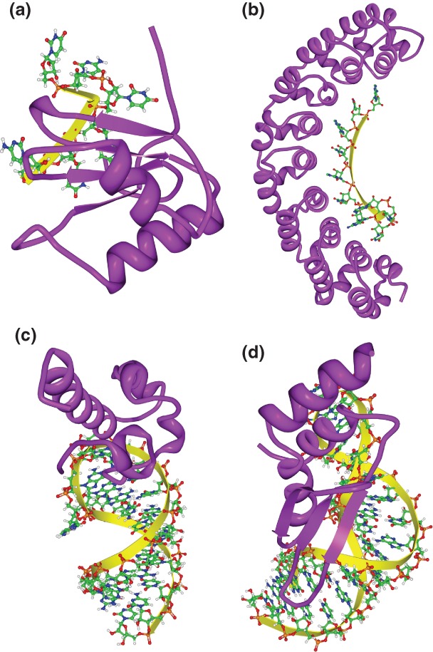
RNA-protein interactions differ from DNA-protein interactions because of the central role of RNA secondary structure. Some RNA-binding domains (RBDs) recognize their target sites mainly by their shape and geometry and others are sequence-specific but are sensitive to secondary structure context. A number of small- and large-scale experimental approaches have been developed to measure RNAs associated in vitro and in vivo with RNA-binding proteins (RBPs). Generalizing outside of the experimental conditions tested by these assays requires computational motif finding. Often RBP motif finding is done by adapting DNA motif finding methods; but modeling secondary structure context leads to better recovery of RBP-binding preferences. Genome-wide assessment of mRNA secondary structure has recently become possible, but these data must be combined with computational predictions of secondary structure before they add value in predicting in vivo binding. There are two main approaches to incorporating structural information into motif models: supplementing primary sequence motif models with preferred secondary structure contexts (e.g., MEMERIS and RNAcontext) and directly modeling secondary structure recognized by the RBP using stochastic context-free grammars (e.g., CMfinder and RNApromo). The former better reconstruct known binding preferences for sequence-specific RBPs but are not suitable for modeling RBPs that recognize shape and geometry of RNAs. Future work in RBP motif finding should incorporate interactions between multiple RBDs and multiple RBPs in binding to RNA.


Figures

Figure 1
Figure 1
Three-dimensional structures of RNA-binding domain (RBD)–RNA complexes. (a) Solution structure of polypyrimidine tract binding (PTB) protein RBD1 in complex with CUCUCU RNA [Protein Data Bank (PDB): 2AD9]. PTB RBD1 binds a YCU site (Y indicating pyrimidine) through β4, β1, and β2, respectively. (b) Co-crystal structure of the PUM-homology domain (PUM-HD) in human Pum1 complexed with a 10-nucleotide single-stranded RNA, 5′-AUUGUACAUA where the last eight nucleotides (UGUACAUA) are individually recognized by three conserved amino acids in Puf repeats 8 to 1, respectively (PDB: 1M8Y). (c) Solution structure of the Vts1p sterile-α motif (specific affinity matrix, SAM) domain in complex with a 5′-CUGGC-3′ pentaloop as part of a 19nt hairpin (PDB: 2ESE). The specific interaction between the Vts1p SAM domain and the target RNA is stabilized by both the direct interaction to the third guanosine base in the RNA pentaloop and the contacts to the unique backbone structure.– (d) Solution structure of dsRBD of yeast Rnt1p in complex with the 5′ terminal AGNN tetraloop of snR47 precursor RNA (PDB: 1T4l). Neither A nor G are recognized by specific hydrogen bonds; instead, the N-terminal helix of the Rnt1p dsRBD interacts with the backbone and the two nonconserved tetraloop bases, by snugly fitting into the minor groove side of the RNA tetraloop and extending into the minor groove at the top of the stem.
Figure 2
Figure 2
Target site accessibility predicts in vivo binding for a diverse range of RNA-binding proteins (RBPs). Comparison of accuracy in predicting bound transcripts based on a given consensus, using either #ATS (i.e., the expected number of accessible target sites, y-axis) or #TS (i.e., the number of target sites, x-axis). Each dot represents the results of an RBP coupled with its previously defined consensus sequence. If there are multiple reported consensus sequences for a protein, the result for each is shown and is distinguished from others by a superscript. Cartoons indicate the species of origin (yeast, fly, or human). RBPs in bold have significantly improved AUROC for #ATS versus #TS (P < 0.05, Delong-Delong-Clarke-Pearson test). The RBDs housed in the RBPs (using SMART domains) are summarized in the pie graph.
Figure 3
Figure 3
Structural context of target sites improves prediction of target mRNAs bound in vivo by RNA-binding proteins (RBPs). Bar graphs compare the accuracy of different methods that use the structural context of motif matches to predict in vivo binding of RBPs. The inset describes the different bars within the graph.
Figure 4
Figure 4
Comparison of prediction accuracy for in vivo binding of nine yeast RNA-binding proteins (RBPs) using parallel analysis of RNA structure (PARS) and RNAplfold to estimate the secondary structure of bound versus unbound transcripts. The results using PARS are shown on the y-axis, those using RNAplfold on the x-axis. (a) The analysis was performed on all consensus sites containing at least one nucleotide with a nonzero PARS score. (b) The analysis was performed only considering nucleotides with nonzero PARS score. (c) As for (b) but with the additional constraint that the transcript load (i.e., reads/nucleotide) was at least five. P-values were calculated using the two-tailed sign test.
Figure 5
Figure 5
RNAcontext-predicted motifs. The figure shows motifs and their structural contexts predicted by RNAcontext using RNAcompete binding data. (Reprinted with permission from Ref . Copyright 2010, PLoS Computational Biology Creative Commons Attribution License.)
Figure 6
Figure 6
Three-dimensional structures of multiple RNA-binding domains (RBDs) in complex with RNA. (a) Solution structure of polypyrimidine tract binding (PTB), RBD3, and RBD4 in complex with CUCUCU RNA [Protein Data Bank (PDB): 2ADC]. RBD3 and RBD4 have different binding specificity: RBD3 binds YCUNN and RBD4 binds YCN (Y, pyrimidine; N, any nucleotide). RBD3 and RBD4 interact extensively, resulting in an antiparallel orientation of their bound RNAs, suggesting that the only way to make these two RBDs bind to a single RNA is to separate their sites by a linker sequence. (b) Solution structure of ADAR2 dsRBD1 and dsRBD2 in complex with GluR-2 R/G RNA (PDB: 2L3J). The dsRBDs recognize their targets by the shape and by the primary sequence in the minor groove. Sequence-specific recognition is achieved through a hydrogen bond to the amino group of G (in the GG mismatch for dsRBD1; in the GC pair for dsRBD2) via a β1-β2 loop and via a hydrophobic contact to adenine H2 (in the AU pair for dsRBD1; in the AC mismatch for dsRBD2) via helix α1. The two dsRBDs bind one face of the RNA and cover about 120° of the turn of the RNA helix.

References

    1. Hogan DJ, Riordan DP, Gerber AP, Herschlag D, Brown PO. Diverse RNA-binding proteins interact with functionally related sets of RNAs, suggesting an extensive regulatory system. PLoS Biol. 2008;6:e255. - PMC - PubMed
    1. Blanchette M, Green RE, MacArthur S, Brooks AN, Brenner SE, Eisen MB, Rio DC. Genome-wide analysis of alternative pre-mRNA splicing and RNA-binding specificities of the Drosophila hnRNP A/B family members. Mol Cell. 2009;33:438–449. - PMC - PubMed
    1. Lukong KE, Chang KW, Khandjian EW, Richard S. RNA-binding proteins in human genetic disease. Trends Genet. 2008;24:416–425. - PubMed
    1. Keene JD. RNA regulons: coordination of post-transcriptional events. Nat Rev Genet. 2007;8:533–543. - PubMed
    1. Seeman NC, Rosenberg JM, Suddath FL, Kim JJ, Rich A. RNA double-helical fragments at atomic resolution. I. The crystal and molecular structure of sodium adenylyl-3′,5′-uridine hexahydrate. J Mol Biol. 1976;104:109–144. - PubMed

FURTHER READING

    1. Messias AC, Sattler M. Structural basis of single-stranded RNA recognition. Acc Chem Res. 2004;37:279–287. - PubMed
    1. Chang KY, Ramos A. The double-stranded RNA-binding motif a versatile macromolecular docking platform. FEBS J. 2005;272:2109–2117. - PubMed
    1. Auweter SD, Oberstrass FC, Allain FH. Sequence-specific binding of single-stranded RNA: is there a code for recognition? Nucleic Acids Res. 2006;34:4943–4959. - PMC - PubMed
    1. Fierro-Monti I, Mathews MB. Proteins binding to duplexed RNA: one motif, multiple functions. Trends Biochem Sci. 2000;25:241–246. - PubMed

Publication types

LinkOut - more resources