. 2014 Jan 15;20(2):331-43.
doi: 10.1158/1078-0432.CCR-13-1736. Epub 2013 Nov 11.

Tumor-reactive CD8+ T cells in metastatic gastrointestinal cancer refractory to chemotherapy

Affiliations

Tumor-reactive CD8+ T cells in metastatic gastrointestinal cancer refractory to chemotherapy

Simon Turcotte et al. Clin Cancer Res. .

Abstract

Purpose: To evaluate whether patients with metastatic gastrointestinal adenocarcinomas refractory to chemotherapy harbor tumor-reactive cytotoxic T cells.

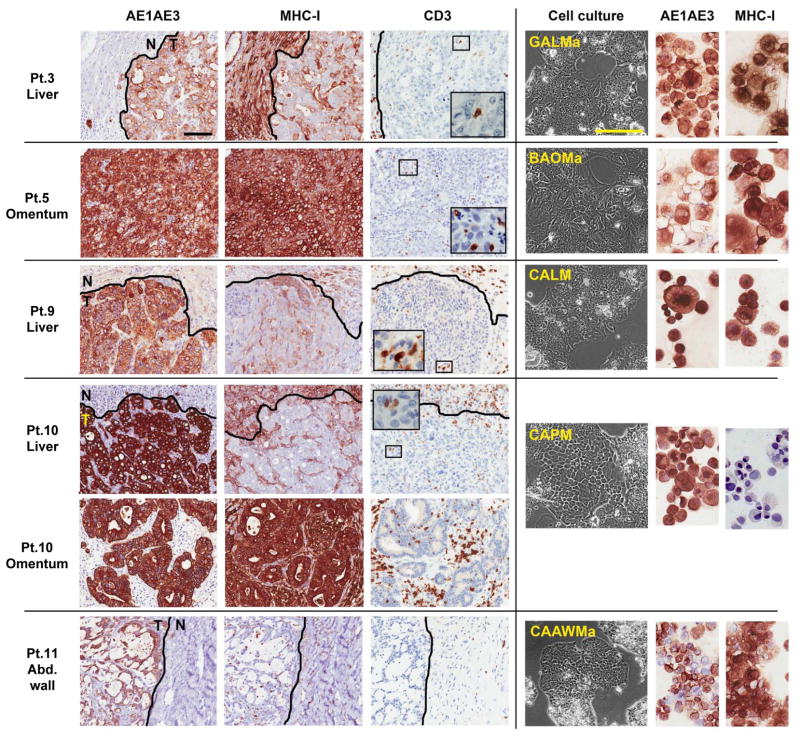
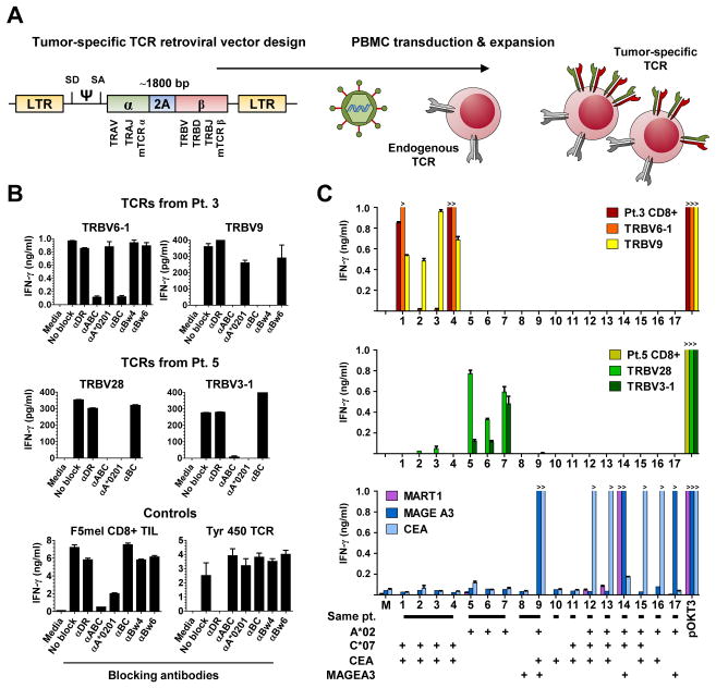
Experimental design: Expansion of CD8(+) tumor-infiltrating lymphocytes (TIL) and cancer cell lines was attempted from gastrointestinal cancer metastases in 16 consecutive patients for the study of antitumor immune recognition. Retroviral transduction of genes encoding T-cell receptors (TCR) was used to define HLA-restriction elements and specific reactivity.

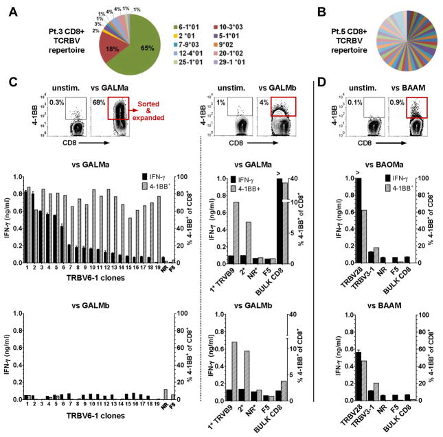
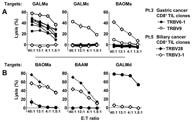
Results: TIL were expanded from metastases in all patients, and new tumor cell lines were generated in 5 patients. Autologous tumor recognition without cross-reactivity against allogeneic HLA-matched gastrointestinal tumors was found in CD8(+) TIL from 3 of these 5 patients. In a patient with gastric cancer liver metastases, the repertoire of CD8(+) TIL was dominated by cytolytic sister clones reactive to 2 out of 4 autologous cancer cell lines restricted by HLA-C*0701. From the same patient, a rare CD8(+) TIL clone with a distinct TCR recognized all four cancer cell lines restricted by HLA-B*4901. In a patient with bile duct cancer, two distinct antitumor cytolytic clones were isolated from a highly polyclonal CD8(+) TIL repertoire. TCRs isolated from these clones recognized epitopes restricted by HLA-A*0201. In a third patient, CD8(+) TIL reactivity was progressively lost against an autologous colon cancer cell line that displayed loss of HLA haplotype.

Conclusions: This study provides a basis for the development of immunotherapy for patients with advanced gastrointestinal malignancies by first establishing the presence of naturally occurring tumor-reactive CD8(+) TIL at the molecular level.


Conflict of interest statement

Conflicts of interests: none

Figures

Fig. 1
Fig. 1. Cancer cell lines established from gastrointestinal cancer metastases with heterogeneous MHC-I expression and weak T-cell infiltration
Immunohistochemical staining of paraffin-embedded gastrointestinal cancer metastases (left) in five patients (Pt.) and representative corresponding cancer cell lines (right). Pt. 3, gastric adenocarcinoma liver metastasis, adjacent normal liver, and derived cancer cell line (GALMa). Pt. 5, omental metastasis from an intrahepatic bile duct adenocarcinoma and derived cancer cell line (BAOMa). Pt. 9, colon adenocarcinoma liver metastasis, adjacent liver, and derived cancer cell line (CALM). Patient 10, colon adenocarcinoma liver metastasis, adjacent liver, and omental metastasis, with derived cancer cell line (CAPM). Patient 11, abdominal wall metastasis from a mucinous colon adenocarcinoma, adjacent stroma, and derived cancer cell line (CAAWMa). All epithelial derived-cancer cells are stained by the pan-cytokeratin marker AE1AE3 which delineate tumor (T) from the adjacent normal tissues (N). MHC class I stains all nucleated cells, with weak and heterogeneous expression found in liver metastases in vivo, however most derived cancer cell line express MHC class I in culture except CAPM. Tumor-infiltrating lymphocytes are revealed by CD3 and T-cell marker, and in all cases represent less than 5% of cells in tumor mass. Representative fields selected from whole slide scan, measure bar = 100 um.
Fig. 2
Fig. 2. Reactivity CD8+ TIL clones to autologous gastric and bile duct cancers
(A) Diversity of the TCR repertoire of CD8+ TIL expanded from patient (Pt.) 3 liver metastasis, where distinct TCR variable β chains sequences are labeled by their TCR germline V consensus regions (TRBV). (B) Highly polyclonal TCRBV repertoire of TIL found in malignant ascites from a bile duct cancer in pt. 5. (C) For Pt. 3, FACS plot of 4-1BB expression upregulation in bulk CD8+ TIL after stimulation by autologous GALMa that allowed enrichment of the 4-1BB+ cells for limiting dilution cloning (red rectangle). Inferior to the FACS plot, the bar graphs represent the reactivity of 19 CD8+ TRBV6-1 TIL clones to the autologous GALMa, but not to the autologous GALMb cancer cell line, a non reactive (NR) clone (TRBV12-4), and the melanoma (F5) CD8+ TIL specificity control. FACS plot (middle) of the ~4 % 4-1BB upregulation (red rectangle) seen in bulk CD8+ TIL from pt.3 after stimulation by autologous GALMb. The bar graphs on the right isde of the dotted line represent the low reactivity found in 2 expanded clones (1*:TRBV9, 2*:unsequenced) to GALMa and GALMb, the bulk CD8+ TIL, a non-reactive (NR) clone, and the F5 specificity control by 4-1BB upregulation; none of these clones secreted significant amount of IFN-γ. (D) For Pt.5, FACS plot of the ~1% 4-1BB upregulation seen in the bulk CD8+ TIL. Limiting dilution cloning allowed isolation of 2 clones (TRBV28 and TRBV3-1) reactive to two autologous cancer cell lines established from an omental metastasis BAOMa and the malignant ascites BAAM. For bar graphs, left axis IFN-γ ng/ml; right axis %4-1BB+ of CD3+CD8+.
Fig. 3
Fig. 3. Specific CD8+ TIL clone lysis of autologous gastric and a bile duct cancer cell lines
Chromium 51-release assays testing cancer cell line lysis by the TRBV6-1 and TRBV9 CD8+ TIL clones from patient 3, and TRBV28 and TRBV3-1 clones from patient 5 in different experiments. (A) Patient 3 TRBV9 CD8+ TIL clone lysed autologous GALMa and GALMc, whereas the TRBV6-1 sister CD8+ clones only lysed autologous GALMa and GALMd (in B). (B) From patient 5, TRBV28 and TRBV3-1 CD8+ TIL clones lysed two autologous cancer cell lines (BAOMa and BAAM). For all clones, reactivity was specific to autologous tumor cell lines.
Fig. 4
Fig. 4. Retroviral transduction of lymphocytes with four TCRs and assessment of their HLA restriction elements and specificity
(A) Design of gammaretroviral TCR construct used for transduction of peripheral blood mononuclear cells (PBMC) with four TCRs derived from patient 3 and 5 cytolytic TIL clones. The human constant chains were replaced with mouse constant chains (mTCRα and mTCRβ) to reduce mispairing of transduced alpha and beta chains with endogenous TCR chains. A ribosomal skipping motif (2A) is inserted between the alpha and beta chains for separate transcription and expression of the two TCR chains. (B) Determination of the restriction elements by which TCR react to autologous cancers (See Table S1 for HLA listing). For TCRs in patient 3, TRBV6-1 TCR recognizes its epitope by HLA-C*0701, whereas TRBV9 is restricted by HLA-B*4901, blocked by Bw4 and not by Bw6, the latter having the potential to block HLA-B*0801. The reactivity of both TCRs in patient 5 are not blocked by the anti-BC antibody, and are restricted by HLA-A*0201. The specificity of the Class I and Class II blocking antibodies was demonstrated in the same experiment by the selective block of known Class I (F5 mel) and Class II (Tyr 450) lymphocyte lines. (C) Reactivity of native TIL and PBMC transduced with TCRs from patient 3 (upper), TCRs from patient 5 (middle), TCR that recognize MART1, MAGE A3 and CEA presented by HLA-A*020101, to 17 targets (see Table S2 for all target tested): 1. GALMa, 2. GALMb, 3. GALMc, 4. GALMd, 5. BAOMa, 6. BAOMb, 7. BAAM, 8. CALM, 9. CALM transduced to express HLA-A*020101, 10. CAPM, 11. CAAWMa, 12. NCI H508 colon cancer (Ca.), 13. SK-CO-1 colon Ca., 14. 624 melanoma TC, 15. SW1463 rectal Ca, 16. KATO III gastric Ca, 17. HCT15 colon Ca. Patients 3 and 5 TCRs reactivity are restricted to the autologous setting.

References

    1. Jemal A, Bray F, Center MM, Ferlay J, Ward E, Forman D. Global cancer statistics. CA Cancer J Clin. 2011;61:69–90. - PubMed
    1. Brahmer JR, Drake CG, Wollner I, Powderly JD, Picus J, Sharfman WH, et al. Phase I study of single-agent anti-programmed death-1 (MDX-1106) in refractory solid tumors: safety, clinical activity, pharmacodynamics, and immunologic correlates. J Clin Oncol. 2010;28:3167–75. - PMC - PubMed
    1. Chung KY, Gore I, Fong L, Venook A, Beck SB, Dorazio P, et al. Phase II study of the anti-cytotoxic T-lymphocyte-associated antigen 4 monoclonal antibody, tremelimumab, in patients with refractory metastatic colorectal cancer. J Clin Oncol. 2010;28:3485–90. - PubMed
    1. Rosenberg SA, Lotze MT, Yang JC, Aebersold PM, Linehan WM, Seipp CA, et al. Experience with the use of high-dose interleukin-2 in the treatment of 652 cancer patients. Ann Surg. 1989;210:474–84. - PMC - PubMed
    1. Royal RE, Levy C, Turner K, Mathur A, Hughes M, Kammula US, et al. Phase 2 trial of single agent Ipilimumab (anti-CTLA-4) for locally advanced or metastatic pancreatic adenocarcinoma. J Immunother. 2010;33:828–33. - PMC - PubMed

Publication types

MeSH terms

Substances