

Repair of UV photolesions in xeroderma pigmentosum group C cells induced by translational readthrough of premature termination codons
- PMID: 24218596
- PMCID: PMC3845163
- DOI: 10.1073/pnas.1312088110
Repair of UV photolesions in xeroderma pigmentosum group C cells induced by translational readthrough of premature termination codons
Abstract
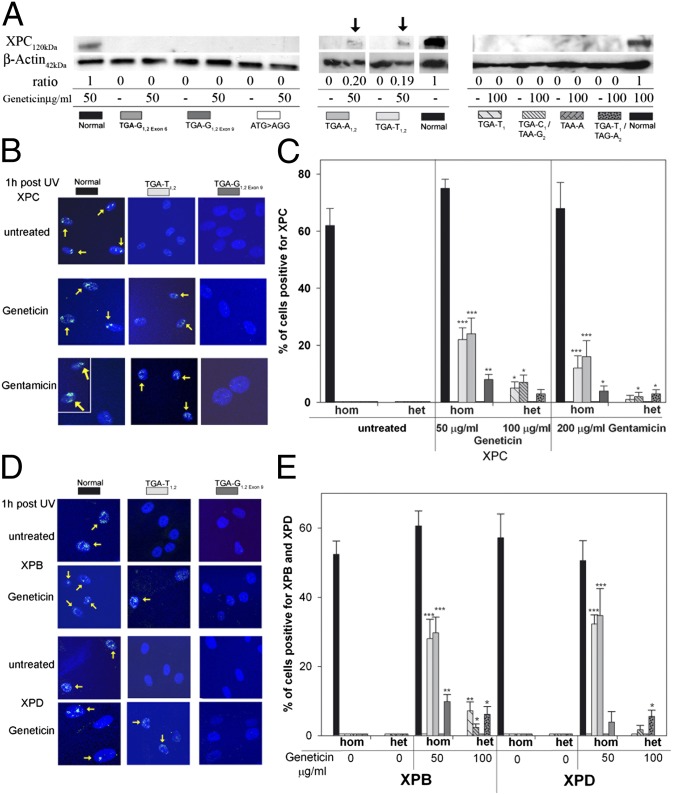
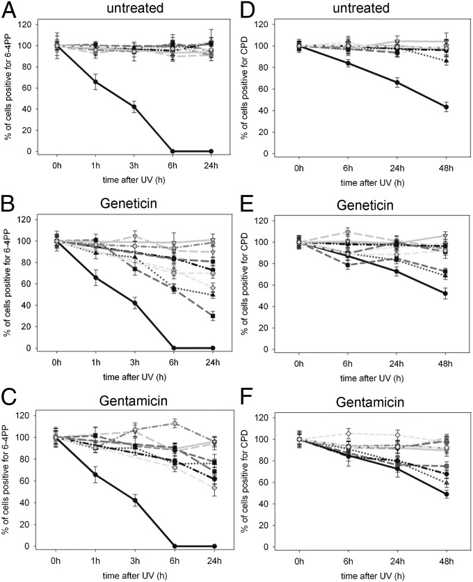
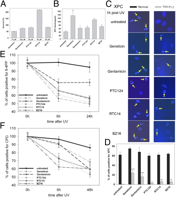
About 12% of human genetic disorders involve premature termination codons (PTCs). Aminoglycoside antibiotics have been proposed for restoring full-length proteins by readthrough of PTC. To assess the efficiency of readthrough, we selected homozygous and compound heterozygous skin fibroblasts from xeroderma pigmentosum (XP) patients with different PTCs in the XPC DNA repair gene. XP patients have a nucleotide excision repair defect and a 10,000-fold increased risk of UV-induced skin cancer. In six of eight PTC-containing XP-C cells, treatment with Geneticin and gentamicin resulted in (i) stabilized XPC-mRNA, which would have been degraded by nonsense-mediated decay; (ii) increased expression of XPC protein that localized to UV-damaged sites; (iii) recruitment of XPB and XPD proteins to UV DNA damage sites; and (iv) increased repair of 6-4 photoproducts and cyclobutane pyrimidine dimers. Expression of PTC in a transfected vector revealed that readthrough depends on the PTC sequence and its location within the gene. This sensitive DNA repair assay system demonstrates the complexity of response to PTC readthrough inducers. The efficiency of aminoglycoside-mediated readthrough depends on the type and copy number of PTC, the downstream 4+ nucleotide, and the location within the exon. Treatment with small-molecule nonaminoglycoside compounds (PTC124, BZ16, or RTC14) resulted in similarly increased XPC mRNA expression and photoproduct removal with less toxicity than with the aminoglycosides. Characterizing PTC structure and parameters governing effective PTC readthrough may provide a unique prophylactic therapy for skin cancer prevention in XP-C patients.
Keywords: UV radiation; readthrough compounds.
Conflict of interest statement
The authors declare no conflict of interest.
Figures
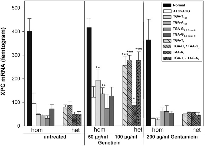
 Normal;
  ATG > AGG;
  TGA-T1,2;
  TGA-T1;
  TGA-A1,2;
  TGA-G1,2 Exon 6;
  TGA-C1/TAA-G2;
  TAA-A1; TGA-G1,2 Exon 9;
  TGA-T1/TAG-A2.
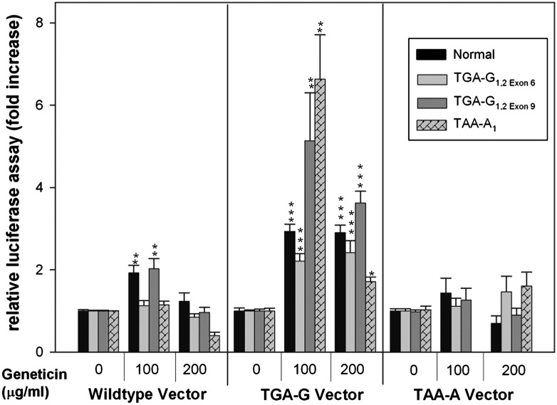
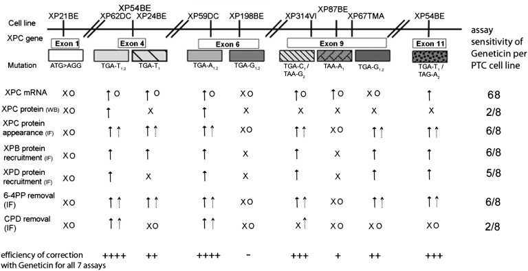
 , Geneticin response;
  , gentamicin response; x, no response to Geneticin; O, no response to gentamicin.
References
-
- Kleijer WJ, et al. Incidence of DNA repair deficiency disorders in western Europe: Xeroderma pigmentosum, Cockayne syndrome and trichothiodystrophy. DNA Repair (Amst) 2008;7(5):744–750. - PubMed
-
- Clement FC, et al. Dynamic two-stage mechanism of versatile DNA damage recognition by xeroderma pigmentosum group C protein. Mutat Res. 2010;685(1-2):21–28. - PubMed
-
- Gozukara EM, et al. A stop codon in xeroderma pigmentosum group C families in Turkey and Italy: Molecular genetic evidence for a common ancestor. J Invest Dermatol. 2001;117(2):197–204. - PubMed
Publication types
MeSH terms
Substances
Grants and funding
LinkOut - more resources
Full Text Sources
Other Literature Sources
Medical
Research Materials
