. 2013 Nov 26;110(48):19360-5.
doi: 10.1073/pnas.1304670110. Epub 2013 Nov 11.

Structural and functional insights into the regulation mechanism of CK2 by IP6 and the intrinsically disordered protein Nopp140

Affiliations

Structural and functional insights into the regulation mechanism of CK2 by IP6 and the intrinsically disordered protein Nopp140

Won-Kyu Lee et al. Proc Natl Acad Sci U S A. .

Abstract

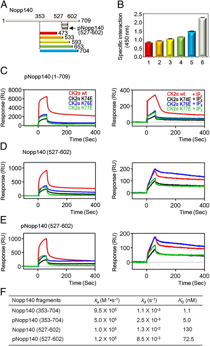
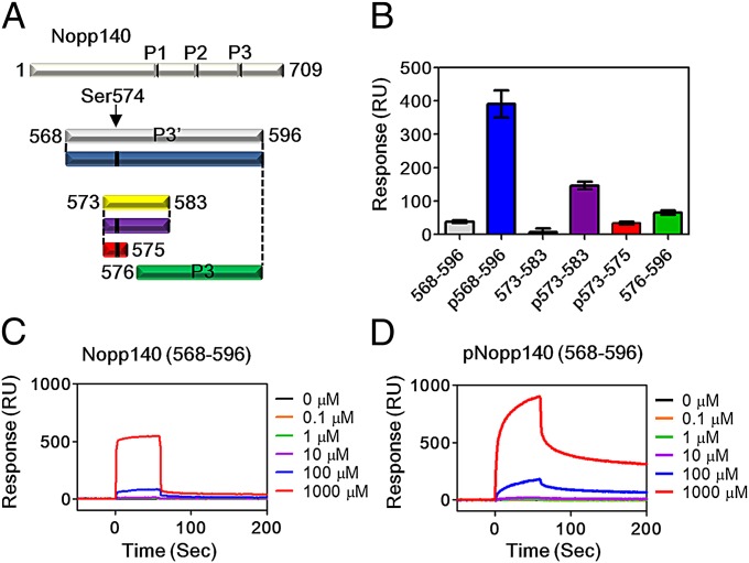
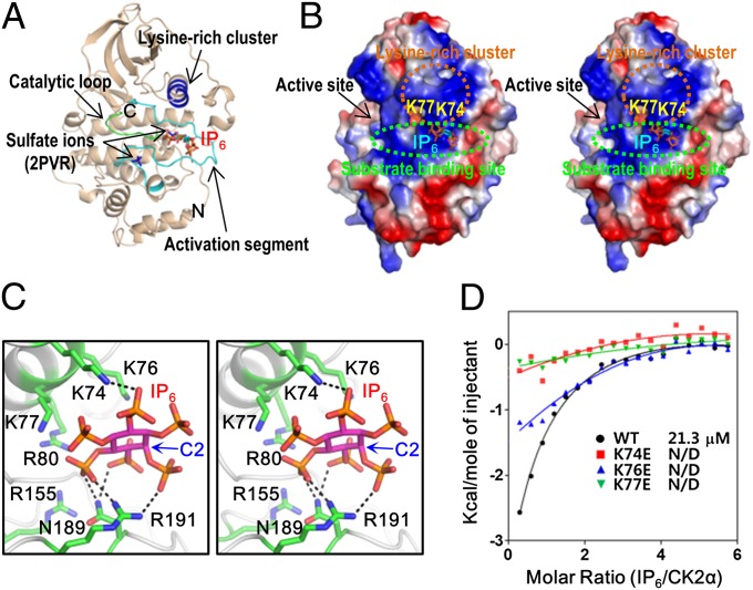
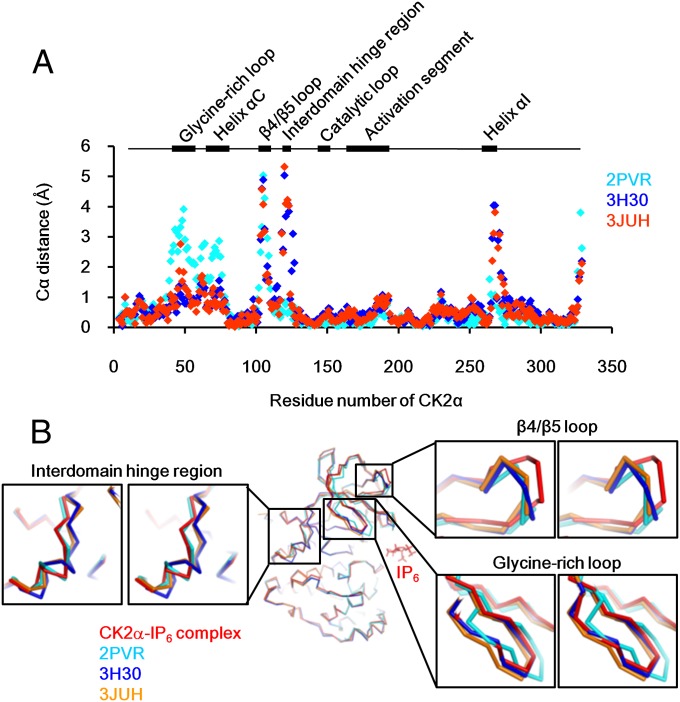
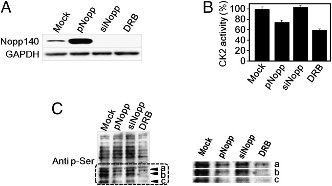
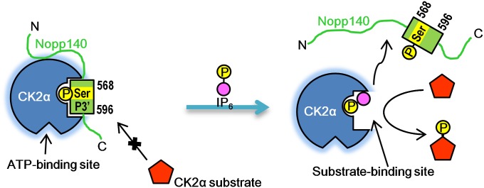
Protein kinase CK2 is a ubiquitous kinase that can phosphorylate hundreds of cellular proteins and plays important roles in cell growth and development. Deregulation of CK2 is related to a variety of human cancers, and CK2 is regarded as a suppressor of apoptosis; therefore, it is a target of anticancer therapy. Nucleolar phosphoprotein 140 (Nopp140), which is an intrinsically disordered protein, interacts with CK2 and inhibits the latter's catalytic activity in vitro. Interestingly, the catalytic activity of CK2 is recovered in the presence of d-myo-inositol 1,2,3,4,5,6-hexakisphosphate (IP6). IP6 is widely distributed in animal cells, but the molecular mechanisms that govern its cellular functions in animal cells have not been completely elucidated. In this study, the crystal structure of CK2 in complex with IP6 showed that the lysine-rich cluster of CK2 plays an important role in binding to IP6. The biochemical experiments revealed that a Nopp140 fragment (residues 568-596) and IP6 competitively bind to the catalytic subunit of CK2 (CK2α), and phospho-Ser574 of Nopp140 significantly enhances its interaction with CK2α. Substitutions of K74E, K76E, and K77E in CK2α significantly reduced the interactions of CK2α with both IP6 and the Nopp140-derived peptide. Our study gives an insight into the regulation of CK2. In particular, our work suggests that CK2 activity is inhibited by Nopp140 and reactivated by IP6 by competitive binding at the substrate recognition site of CK2.

Keywords: IDP; inositol hexakisphosphate; phosphorylation.


Conflict of interest statement

The authors declare no conflict of interest.

Figures

Fig. 1.
Fig. 1.
Mapping binding region of Nopp140 with CK2α and the inhibitory effect of IP6. (A) Schematic representation of Nopp140 constructs. (B) Specific interaction between C-terminal domain of Nopp140 and CK2α observed using a modified ELISA method. Purified CK2α was immobilized on a plate, and the intensities of the chemiluminescence signal from the bound HRP-conjugated anti–His-tag antibody with His-Nopp140 fragments, 353–473 (red), 353–533 (orange), 353–593 (yellow), 353–653 (green), 353–704 (cyan), and 1–709 (white) were measured. (C) SPR sensorgrams of pNopp140 (1–709) with CK2α, CK2α wild type (colored in red), and CK2α mutants (K74E in black, K76E in blue, and K77E in green) were used as analytes with (Right) and without IP6 (Left). (D) SPR sensorgrams of Nopp140 (527–602) with CK2α. The analytes were used as indicated in Fig. 1C. (E) SPR sensorgrams of pNopp140 (527–602) with CK2α. The analytes were used as indicated in Fig. 1C. (F) Kinetic data for the interaction between CK2α wild-type and Nopp140 fragments. Original SPR sensorgrams are shown in Fig. S3.
Fig. 2.
Fig. 2.
Analysis of the narrow interaction region of Nopp140 and CK2α. (A) Schematic representation of full-length Nopp140, three Nopp140 peptides (P1, P2, and P3), and six P3′-derived peptides (colored in gray, blue, yellow, purple, red, and green). The position and phosphorylation of Ser574 is indicated by an arrow and black bar, respectively. (B) SPR analysis of the interaction between P3′-derived Nopp140 peptides and CK2α. Nopp140 (568–596), pNopp140 (568–596), Nopp140 (573–583), pNopp140 (573–583), Nopp140 (573–575), and Nopp140 (576–596) are represented by gray, blue, yellow, purple, red, and green columns, respectively. SPR sensorgrams of CK2α with (C) Nopp140 (568–596) and (D) pNopp140 (568–596) were obtained for 0.1, 1, 10, 100, and 1,000 μM Nopp140 peptides, respectively.
Fig. 3.
Fig. 3.
Crystal structure of CK2α in complex with IP6. (A) Ribbon diagram of the CK2α–IP6 complex. The positions of the activation segment, catalytic loop, and lysine-rich cluster are indicated. Sulfate ions are incorporated from the structure of CK2α in complex with sulfate ions (PDB ID code 2PVR). (B) Stereoview of the electrostatic potential at the molecular surface of CK2α. IP6 is represented by a stick diagram. (C) A close-up stereoview of IP6 binding to CK2α. The black dotted lines denote hydrogen bonds. (D) ITC of IP6 into CK2α solution. The wild-type CK2α and its mutants (K74E, K76E, and K77E) were titrated against IP6. Original ITC data are shown in Fig. S5. N/D denotes the dissociation constant (KD) is not determined.
Fig. 4.
Fig. 4.
Conformations of variable regions of CK2α. (A) Distance plots between the Cα position of the CK2α–IP6 complex and the corresponding Cα positions of various CK2α structures (PDB ID codes 2PVR, 3H30, and 3JUH colored in cyan, blue, and orange, respectively). The rectangles above the plots represent the positions of variable regions of CK2α. (B) Stereoview of superposition of the CK2α–IP6 complex (red) with various CK2α structures (PDB ID codes 2PVR, 3H30, and 3JUH colored in cyan, blue, and orange, respectively).
Fig. 5.
Fig. 5.
In vivo inhibitory effect of Nopp140 on CK2. (A) Overexpression or knockdown of Nopp140. HeLa cells were transfected with the Nopp140 expression vector (pNopp) or siRNAs targeting Nopp140 (siNopp). For the Western blot, 20 μg of the proteins from the cleared lysates were used. (B) Negatively regulated CK2 activity by Nopp140. The cleared lysates of the HeLa cells transfected with pNopp or siNopp were incubated with α-casein. The reaction was further reacted with Kinase-Glo, and the luminescence intensity was measured using a microplate luminometer. The CK2 inhibitor DRB was used as a reference under the same conditions. All experiments were performed in triplicate. (C) Selectively regulated CK2 activity by Nopp140. The lysates of the HeLa cells treated with DRB or transfected with pNopp140 or siNopp140 were immunoblotted with phosphoserine antibodies.
Fig. 6.
Fig. 6.
Proposed model of CK2α regulation by IP6 and Nopp140.

References

    1. Lee HH, Elia N, Ghirlando R, Lippincott-Schwartz J, Hurley JH. Midbody targeting of the ESCRT machinery by a noncanonical coiled coil in CEP55. Science. 2008;322(5901):576–580. - PMC - PubMed
    1. Tawfic S, et al. Protein kinase CK2 signal in neoplasia. Histol Histopathol. 2001;16(2):573–582. - PubMed
    1. Seldin DC, Leder P. Casein kinase II alpha transgene-induced murine lymphoma: Relation to theileriosis in cattle. Science. 1995;267(5199):894–897. - PubMed
    1. Ahmed K, et al. Significance of protein kinase CK2 nuclear signaling in neoplasia. J Cell Biochem Suppl. 2000;35(Suppl 35):130–135. - PubMed
    1. St-Denis NA, Litchfield DW. Protein kinase CK2 in health and disease: From birth to death: The role of protein kinase CK2 in the regulation of cell proliferation and survival. Cell Mol Life Sci. 2009;66(11-12):1817–1829. - PMC - PubMed

Publication types

Associated data

LinkOut - more resources