. 2014 Jan 3;289(1):476-84.
doi: 10.1074/jbc.M113.504472. Epub 2013 Nov 12.

Nitric oxide activation of guanylate cyclase pushes the α1 signaling helix and the β1 heme-binding domain closer to the substrate-binding site

Affiliations

Nitric oxide activation of guanylate cyclase pushes the α1 signaling helix and the β1 heme-binding domain closer to the substrate-binding site

Mareike Busker et al. J Biol Chem. .

Abstract

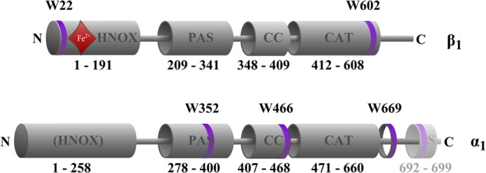
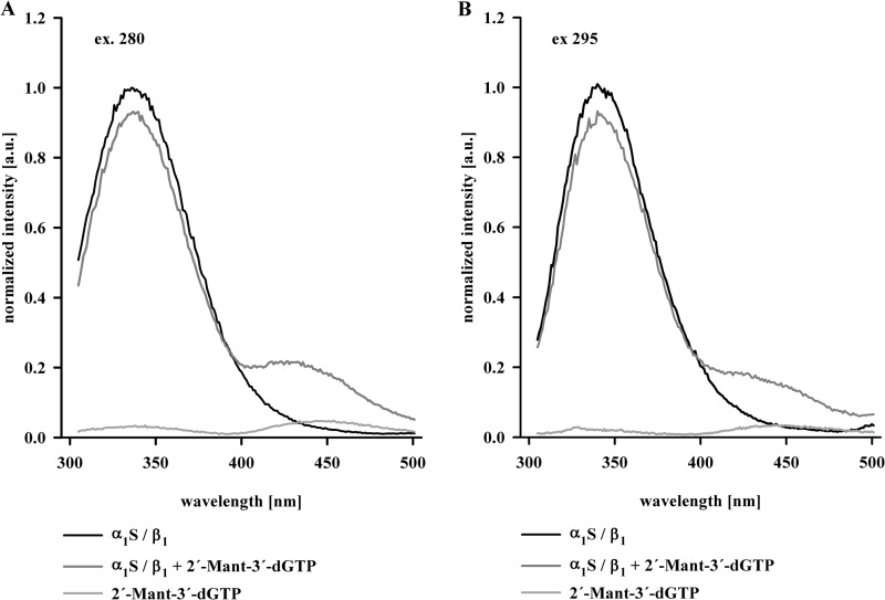
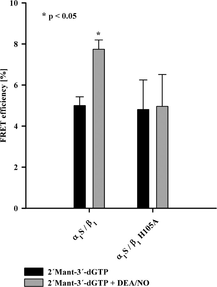
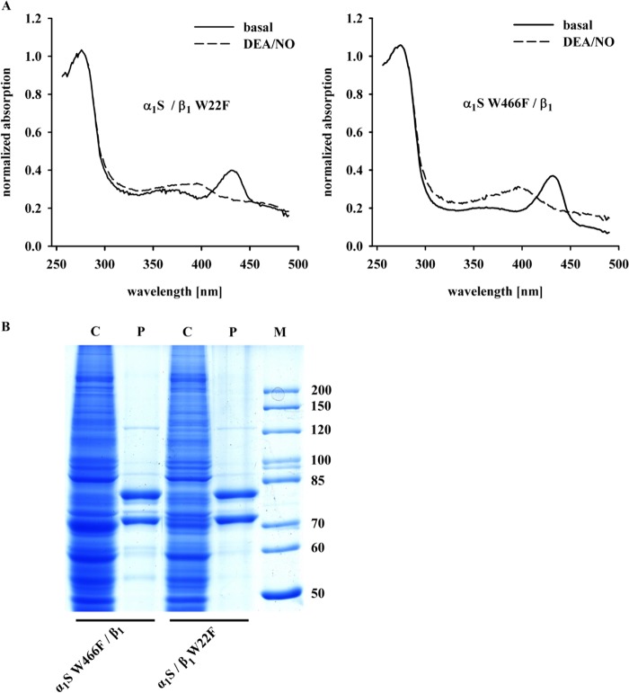
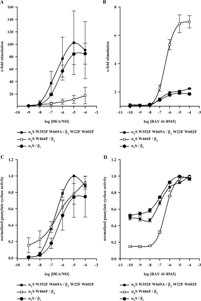
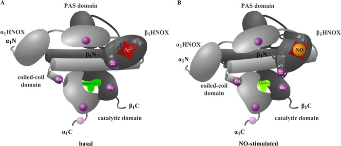
The complete structure of the assembled domains of nitric oxide-sensitive guanylate cyclase (NOsGC) remains to be determined. It is also unknown how binding of NO to heme in guanylate cyclase is communicated to the catalytic domain. In the current study the conformational change of guanylate cyclase on activation by NO was studied using FRET. Endogenous tryptophan residues were used as donors, the substrate analog 2'-Mant-3'-dGTP as acceptor. The enzyme contains five tryptophan residues distributed evenly over all four functional domains. This provides a unique opportunity to detect the movement of the functional domains relative to the substrate-binding catalytic region. FRET measurements indicate that NO brings tryptophan 22 in the αB helix of the β1 heme NO binding domain and tryptophan 466 in the second short helix of the α1 coiled-coil domain closer to the catalytic domain. We propose that the respective domains act as a pair of tongs forcing the catalytic domain into the nitric oxide-activated conformation.

Keywords: Ciguate; Cyclic GMP (cGMP); Fluorescence Resonance Energy Transfer (FRET); Guanylate Cyclase (Guanylyl Cyclase); Nitric Oxide; Signal Transduction.


Figures

FIGURE 1.
FIGURE 1.
Positions of the five tryptophan residues in rat NOsGC subunits. Tryptophan residues are marked as purple rings. Amino acids 692–699 represent the Strep-tag II (S) containing an additional tryptophan residue. The red rhombus indicates position of the heme group. The schematic representation of functional domains is based on the crystallized region of the HNOX domain of Alr2278 from Nostoc sp. (PCC 7120) (32) on the crystallized region of PAS domain of signal transduction histidine kinase from Nostoc punctiforme (PCC 73102) (33) on the crystallized region of the coiled-coil domain of the NOsGC β1 subunit from Rattus norvegicus (24) and on the crystallized catalytic region of the guanylate cyclase CYG12 from Chlamydomonas reinhardtii (34).
FIGURE 2.
FIGURE 2.
Emission of NOsGC in absence and presence of 2′-Mant-3′-dGTP. Normalized emission curves of wild-type NOsGC at 280 nm excitation (A) and at 295 nm excitation (B) in the absence of 2′-Mant-3′-dGTP (black line) and in the presence of 2′-Mant-3′-dGTP (3 μm) (gray line) are presented. For comparison, 2′-Mant-3′-dGTP autofluorescence is shown (light gray line). Graphs show emission curves of one of four representative measurements (a.u., arbitrary unit).
FIGURE 3.
FIGURE 3.
FRET efficiency of NOsGC under basal and NO-stimulated conditions. FRET efficiencies (%) of wild-type NOsGC and NO-insensitive NOsGC mutant at 295 nm excitation in the presence of 2′-Mant-3′-dGTP (black columns) and in the presence of 2′-Mant-3′-dGTP and DEA/NO 100 μm (gray columns) are shown. An asterisk marks p value < 0.05 compared with values in the absence of DEA/NO. The results are expressed as means ± S.E. of at least four independent experiments.
FIGURE 4.
FIGURE 4.
Comparison of FRET efficiencies of NOsGC mutants. A, FRET efficiency (%) under basal conditions for NOsGC wild-type enzyme and NOsGC mutants are compared. B, change in FRET efficiency (%) after the addition of DEA/NO (100 μm) for NOsGC wild-type enzyme and NOsGC mutants. Values were calculated as the difference of basal FRET efficiency to NO-stimulated FRET efficiency. An asterisk marks p value < 0.05 compared with wild-type enzyme. The results are expressed as means ± S.E. of at least three independent experiments.
FIGURE 5.
FIGURE 5.
Absorbance spectra and SDS-PAGE analysis of NOsGC mutants. A, presented are the normalized absorbance spectra for α1S/β1-W22F and α1S W466F/β1 in the absence and presence of DEA/NO (100 μm). Graphs show absorbance spectra of one of three representative measurements. B, Coomassie staining of SDS-PAGE analysis for α1S/β1-W22F and α1S-W466F/β1. C indicates lanes with cytosolic fractions (50 μg), and P marks lanes with purified enzymes (1 μg). The marker M is given in kDa.
FIGURE 6.
FIGURE 6.
Dose-response curves of NOsGC wild-type enzyme and NOsGC mutants. Concentration-dependent activation of α1S/β1, α1S-W352F,W669A/β1-W22F,W602F and α1S-W466F/β1 was measured in a range of 1 nm to 100 μm DEA/NO or 0.1 nm to 100 μm BAY 41-8543 in the presence of DEA/NO (100 μm). A, fold stimulation by DEA/NO. B, fold stimulation by BAY 41-8543 in the presence of DEA/NO. C and D, same data as in A and B normalized to the maximum. The results are expressed as means ± S.E. of at least three independent experiments.
FIGURE 7.
FIGURE 7.
Model of NOsGC signal transmission. The model was developed on basis of NOsGC structure postulated by Fritz et al. (26). The α1 subunit is shown in light gray, and the β1 subunit is shown in dark gray.

References

    1. Misono K. S., Philo J. S., Arakawa T., Ogata C. M., Qiu Y., Ogawa H., Young H. S. (2011) Structure, signaling mechanism and regulation of the natriuretic peptide receptor guanylate cyclase. FEBS J. 278, 1818–1829 - PMC - PubMed
    1. Derbyshire E. R., Marletta M. A. (2012) Structure and regulation of soluble guanylate cyclase. Annu. Rev. Biochem. 81, 533–559 - PubMed
    1. Ogawa H., Qiu Y., Ogata C. M., Misono K. S. (2004) Crystal structure of hormone-bound atrial natriuretic peptide receptor extracellular domain: rotation mechanism for transmembrane signal transduction. J. Biol. Chem. 279, 28625–28631 - PubMed
    1. Padayatti P. S., Pattanaik P., Ma X., van den Akker F. (2004) Structural insights into the regulation and the activation mechanism of mammalian guanylyl cyclases. Pharmacol. Ther. 104, 83–99 - PubMed
    1. Winger J. A., Marletta M. A. (2005) Expression and characterization of the catalytic domains of soluble guanylate cyclase: interaction with the heme domain. Biochemistry 44, 4083–4090 - PubMed

Publication types

MeSH terms

LinkOut - more resources