Review
. 2013 Dec;14(12):775-86.
doi: 10.1038/nrm3699. Epub 2013 Nov 13.

The biophysics and cell biology of lipid droplets

Affiliations
Review

The biophysics and cell biology of lipid droplets

Abdou Rachid Thiam et al. Nat Rev Mol Cell Biol. 2013 Dec.

Abstract

Lipid droplets are intracellular organelles that are found in most cells, where they have fundamental roles in metabolism. They function prominently in storing oil-based reserves of metabolic energy and components of membrane lipids. Lipid droplets are the dispersed phase of an oil-in-water emulsion in the aqueous cytosol of cells, and the importance of basic biophysical principles of emulsions for lipid droplet biology is now being appreciated. Because of their unique architecture, with an interface between the dispersed oil phase and the aqueous cytosol, specific mechanisms underlie their formation, growth and shrinkage. Such mechanisms enable cells to use emulsified oil when the demands for metabolic energy or membrane synthesis change. The regulation of the composition of the phospholipid surfactants at the surface of lipid droplets is crucial for lipid droplet homeostasis and protein targeting to their surfaces.


Figures

Figure 1
Figure 1. Basic principles of emulsion physics relevant to lipid droplets
a. Surface lipids on LDs and their curvatures. Curvature of typical surfactants is defined according to the difference between the area occupied by their hydrophilic head and the area of their lipophilic tail. Positive curvatures correspond to a predominant hydrophilic part. Positively curved lipids, lyso-phospholipids, PI, and MAG, tend proffer a monolayer a positive curvature. Monolayers mainly formed by DAG, PA, or cholesterol on the other hand have a negative curvature. PC is cylindrical in shape and has almost no curvature. PC therefore generally assembles into lamella and is the main component of bilayer membranes. b. Influence of surfactants on emulsion stability. More surfactant at the interface tends to lower surface area A and increase stability. Less surfactant, or less effective surfactants (e.g., PE vs. PC) tend to do the opposite. c. Elasticity of surface monolayers. A loose monolayer, such as an oil-water interface, has wrinkles associated to thermal fluctuations. The presence of phospholipids dampens the fluctuations by creating an energy barrier to surface deformation. They also increase the elasticity of the monolayer. Phospholipids with longer acyl chains are more efficient for dampening fluctuations (left). Likewise, higher concentrations of phospholipids result in a higher barrier to induce deformation by thermal fluctuation (right). The presence of proteins also increases the elasticity. d. Laplace pressure is the pressure that builds up inside the drop to counterbalance the compression effect of surface tension. P0 is pressure in the continuous phase. The surface tension is denoted γ. The drop radius is r. The Laplace pressure of the drop is the difference between the pressures inside and outside the drop and corresponds to 2γ/r. If surface tension is similar, smaller drops have higher Laplace pressures than larger drops
Figure 2
Figure 2. Processes that govern changes in lipid droplet size
a. Coalescence and influence of monolayer curvature. A TG droplet covered with phospholipids forms a pore with another monolayer that can be of another LD or the outer monolayer of a bilayer. At site of the pore, the monolayer is bent, and monolayer curvature, depending on the types of lipids, becomes important. If the spontaneous curvature of the monolayer is positive (e.g., in excess presence of positively curved lipids), this results in a “frustrated” situation, with high line tension, and the pore closes. If the monolayer’s spontaneous curvature is negative, e.g. in excess presence of negatively curved lipids, the curvature of the lipids matches the bending, and the line tension is low. Therefore the pore is stable and can open further. In the case of two LDs, this results in fusion or coalescence, generating one larger LD (inlay). In the case of a LD and a membrane, fusion results in a transiently stable connection of LDs with bilayers. Pore opening and fusion occur in a millisecond scale. b. Ripening of LDs. In ripening, molecules from one LD diffuse to another. The direction is determined by the difference in Laplace pressures of the two LDs, with TG molecules traveling from smaller LDs to bigger LDs. In the case of TGs and LDs, diffusion might occur in swollen micelles, which are micelles containing small amounts of TG. In contrast to coalescence, ripening takes several minutes. The volume increase of the bigger drop is linear over time, r3∞t. Ripening leads also fewer and bigger LDs; however, one droplet shrinks while the other one grows. c. Growth of LDs by new TG synthesis in situ. Enzymes mediating TG synthesis, GPAT4, AGPAT3 and DGAT2, can directly localize to LDs and synthesize TG at the surfaces of LDs. Acyl CoA synthetases localize to LDs and likely provide the fatty acyl CoA substrates. As recently observed , the volume of the drop increases linearly over time, r3∞t.
Figure 3
Figure 3. Binding mode of proteins
Illustration of amphipathic helices and hairpin contained proteins binding to LDs. (left) A loose monolayer of higher surface tension is bound by one type of helix (CCTα or ApoA’s C-terminus for example) and a hairpin (of GPAT4 for example). A helix, typically of ApoE, prefers staying in the cytosolic phase. (right) The compressed monolayer could be still bound by the hairpin. The helix that bound the loose monolayer is now expulsed. The protein free in the cytosol could prefer folding its amphipathic helix by interacting with the head group of phospholipid and bind.
Figure 4
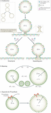
Figure 4. Models for mechanisms of lipid droplet formation
a. Illustration of dewetting transitions. A liquid deposited on a surface can wet completely the surface and form a liquid film on top of it. The liquid can dewet and form a drop with a contact angle with the substrate. Wetting is controlled by surface tension. b. Model for spontaneous budding of a TG droplet from a bilayer based on dewetting transition. Accumulation of TG inside a bilayer can lead to spontaneous emulsification of a TG drop in a low-surface tension environment. The expulsion of the TG LD corresponds to a dewetting state that is energetically favored. Different surface lipids can modulate such process. c. Origins of different LD populations. Evidence suggests that two populations of basic LDs exist. Smaller LDs bud from the ER (e.g., from the enzymatic products of ER enzymes such as DGAT1). Such LDs pathway has a characteristic size that likely depends on the surface surfactants. A second population of cytosolic LDs, called expanding LDs, acquires enzymes of the GPAT4/DGAT2 TG synthesis pathway. These enzymes can locally synthesize TG at the LD from local substrates.
Figure 5
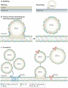
Figure 5. The utilization of consumption of lipid droplets
a. Scheme of lipolysis. ATGL catalyzes the first step of lipolysis and is recruited onto LDs by a co-factor CGI58. TG hydrolysis by ATGL leads to a release of free FA and mainly 1,3-DAG. HSL, also bound to LDs, hydrolyses DAG into FFA and MAG. The latter is in turn hydrolyzed to FFA and glycerol by MGL, which is soluble in the cytosol. The volume of the LD decreases over time, probably linearly. As a result, the surface phospholipids and proteins become more crowded. Mechanisms must exist to facilitate the catabolism or removal of these surface components (discussed in text). b. Scheme of LD autophagy, or lipophagy. Autophagy is proposed to also regulate LDs degradation and TG utilization. An autophagosome forms in the cytosol and encapsulates a LD. It subsequently fuses with a lysosomal organelle containing hydrolytic enzymes that hydrolyze TG from the encapsulated LD and proteases that degrade LD proteins.

References

    1. Walther TC, Farese RV., Jr. Lipid droplets and cellular lipid metabolism. Annu Rev Biochem. 2012;81:687–714. - PMC - PubMed
    1. Brasaemle DL, Wolins NE. Packaging of fat: an evolving model of lipid droplet assembly and expansion. J Biol Chem. 2012;287:2273–9. - PMC - PubMed
    1. Fujimoto T, Parton RG. Not just fat: the structure and function of the lipid droplet. Cold Spring Harb Perspect Biol. 2011;3 - PMC - PubMed
    1. Thiele C, Spandl J. Cell biology of lipid droplets. Curr Opin Cell Biol. 2008;20:378–85. - PubMed
    1. Krahmer N, Farese RV, Jr., Walther TC. Balancing the fat: lipid droplets and human disease. EMBO Mol Med. 2013;5:905–15. - PMC - PubMed

Publication types

Substances