. 2013 Dec 27;288(52):37112-25.
doi: 10.1074/jbc.M113.514398. Epub 2013 Nov 12.

DNA-dependent protein kinase regulates DNA end resection in concert with Mre11-Rad50-Nbs1 (MRN) and ataxia telangiectasia-mutated (ATM)

Affiliations

DNA-dependent protein kinase regulates DNA end resection in concert with Mre11-Rad50-Nbs1 (MRN) and ataxia telangiectasia-mutated (ATM)

Yi Zhou et al. J Biol Chem. .

Abstract

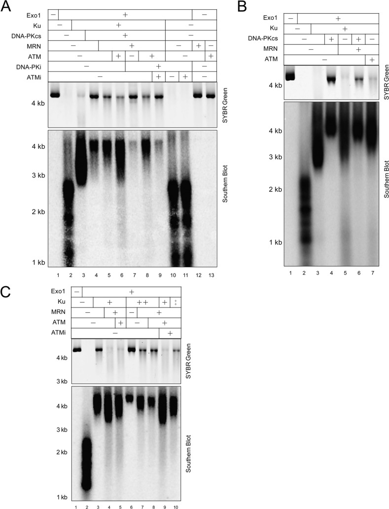
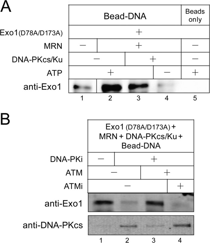
The resection of DNA double strand breaks initiates homologous recombination (HR) and is critical for genomic stability. Using direct measurement of resection in human cells and reconstituted assays of resection with purified proteins in vitro, we show that DNA-dependent protein kinase catalytic subunit (DNA-PKcs), a classic nonhomologous end joining factor, antagonizes double strand break resection by blocking the recruitment of resection enzymes such as exonuclease 1 (Exo1). Autophosphorylation of DNA-PKcs promotes DNA-PKcs dissociation and consequently Exo1 binding. Ataxia telangiectasia-mutated kinase activity can compensate for DNA-PKcs autophosphorylation and promote resection under conditions where DNA-PKcs catalytic activity is inhibited. The Mre11-Rad50-Nbs1 (MRN) complex further stimulates resection in the presence of Ku and DNA-PKcs by recruiting Exo1 and enhancing DNA-PKcs autophosphorylation, and it also inhibits DNA ligase IV/XRCC4-mediated end rejoining. This work suggests that, in addition to its key role in nonhomologous end joining, DNA-PKcs also acts in concert with MRN and ataxia telangiectasia-mutated to regulate resection and thus DNA repair pathway choice.

Keywords: DNA Damage Response; DNA Enzymes; DNA Recombination; DNA Repair; DNA-binding Protein; Protein Kinases.


Figures

FIGURE 1.
FIGURE 1.
Chemical inhibition of DNA-PK stimulates DSB resection in human cells. A, schematic diagram of TaqMan qPCR primer design and in vivo resection assay. Primers for measurement of ssDNA generated by resection at various sites adjacent to the AsiSI-induced DSB1 are indicated by arrows. The primers are designed across restriction sites (here shown as BsrGI) located varying distances from the AsiSI site. Primer design for resection measurement at “DSB2” and No DSB is similar, except that the primer pairs for DSB2 are across BamHI sites, and the primer pair for No DSB is across a HindIII restriction site. Each genomic DNA sample from cells treated or mock-treated with 4-OHT was digested or mock-digested with restriction enzyme (BsrGI, BamHI-HF, or HindIII-HF), followed by qPCR analysis using corresponding set(s) of primers and probes. For each sample, a ΔCt was calculated by subtracting the Ct value of the mock-digested sample from the Ct value of the digested sample, and the percentage of resected DNA was calculated as described under “Experimental Procedures.” B, ER-AsiSI U2OS cells were pretreated with 10 μm ATMi KU-55933 or 10 μm DNA-PK inhibitor NU-7441 (DNA-PKi) for 1 h, followed by induction or mock induction of DSBs with 300 nm 4-OHT for 4 h and measurement of DNA resection. C, percentages of DSBs at the two selected AsiSI sites were measured by qPCR using undigested gDNA samples from B and two sets of primers across the two AsiSI sites. The No DSB primers were used to normalize the amount of gDNA in the qPCR. DSB percentages at DSB1 and DSB2 sites in mock-induced cells were both set to zero. D, ER-AsiSI U2OS cells were pretreated with 10 μm ATMi KU-55933, 10 μm DNA-PK inhibitor NU-7441, or 20 μm DNA-PK inhibitor NU-7026 as indicated for 1 h, followed by induction of DSBs with 300 nm 4-OHT for 4 h. Cell lysates were analyzed by Western blot using antibodies against DNA-PKcs, phospho-DNA-PKcs Ser-2056, ATM, and phospho-ATM Ser-1981, with PARP-1 as a loading control. E, quantitation of DNA-PKcs band in D; average of three quantitations is shown with standard deviation. F, ER-AsiSI U2OS cells were treated as in D, and DNA resection was measured. Error bars show standard deviation (n = 3 or 4). nt, nucleotide.
FIGURE 2.
FIGURE 2.
DNA-PKcs shows an inhibitory effect on DNA end resection by Exo1 in vitro. A, purified recombinant proteins used in this study. Exo1, Ku, DNA-PKcs, wild-type MRN (WT) and nuclease-deficient MRN (H129L/D130V), ATM, and DNA LigIV-XRCC4 were stained with Coomassie Blue after SDS-PAGE. The Ku-associated WT DNA-PKcs and Thr-2609 cluster phospho-blocking DNA-PKcs mutant (6A) were separated by SDS-PAGE followed by silver staining. Asterisk indicates Hsp70 that copurifies with ATM. B, schematic diagram of the resection assay in which a 4.4-kb linear plasmid DNA is incubated with Exo1 and other factors in the reaction, which leads to either no resection, short resection tracks, or medium to long resection tracks (shown with resection initiating from both DNA ends). The reaction products are visualized with SYBR Green (filled circle), which recognizes duplex DNA, or by nondenaturing Southern blot analysis using an RNA probe (line with asterisks) specific for a 1-kb region of the 3′ strand. C, reconstituted DSB resection assay was performed in the presence of 0.5 nm Exo1, 14 nm Ku, 7 nm DNA-PKcs, and 37 pm linear DNA at 37 °C for 1 h. Reaction products were separated by 0.7% native agarose gel and visualized as described in B by SYBR Green staining (top), followed by a nondenaturing Southern blot (bottom). D, DNA resection assays were performed as in C in the presence or absence of ATP. E, DNA resection assays were performed as in C but were stopped after 1 or 2 h as indicated. F, DNA resection assays were performed for 1 or 2 h with 0.35 nm wild-type (WT) DNA-PKcs or a Thr-2609 cluster site phospho-blocking DNA-PKcs mutant (6A) in the presence of 1.9 nm Ku, 0.1 nm Exo1, and 37 pm 4.4 kb linear DNA.
FIGURE 3.
FIGURE 3.
MRN overcomes DNA-PKcs inhibition of Exo1-mediated resection. A, DNA resection assays were performed as in Fig. 2C in the presence of 0.5 nm Exo1, 14 nm Ku, 7 nm DNA-PKcs, 37 pm 4.4-kb linear DNA, but with MRN added as indicated (6, 12, or 24 nm). The 3′ to 5′ exonuclease activity of Mre11 is not active under these reaction conditions (in the absence of manganese) (51). B, DNA resection assays were performed in the presence of 0.35 nm WT DNA-PKcs or a Thr-2609 cluster site phospho-blocking DNA-PKcs mutant (6A), 1.9 nm Ku, 0.1 nm Exo1, and 37 pm 4.4-kb linear DNA with or without 9.5 nm MRN complex for 1 h. C, DNA resection assays as in Fig. 3A were performed with 24 or 72 nm MRN or nuclease-deficient MRN mutant (M(H129L/D130V)RN) in the absence of ATP or in the presence of 200 μm DNA-PKi NU7026. D, part of the DNA products in C were digested or mock-digested with NciI at 37 °C overnight, and resection was measured by qPCR as described under “Experimental Procedures.” A representative experiment is shown. nt, nucleotide.
FIGURE 4.
FIGURE 4.
ATM promotes Exo1-mediated resection in the presence of Ku and DNA-PKcs. A, DNA resection assays were performed as in Fig. 2C in the presence of 0.5 nm Exo1, 14 nm Ku, 7 nm DNA-PKcs, 37 pm 4.4-kb linear DNA, 24 nm MRN, 0.1 nm ATM, 20 μm ATM-specific inhibitor KU-55933, and 200 μm DNA-PK-specific inhibitor NU7026. Reaction products were visualized by SYBR Green staining (top) and Southern blot analysis (bottom). B, DNA resection assays were performed as in A using indicated proteins. C, DNA resection assays were performed as in A with 0.5 nm Exo1, 56 nm (+), or 112 nm (++) Ku, 24 nm MRN, and 0.1 nm ATM in the presence or absence of 20 μm ATM-specific inhibitor KU-55933.
FIGURE 5.
FIGURE 5.
ATM kinase activity compensates for DNA-PKcs autophosphorylation upon chemical inhibition of DNA-PKcs. A, resection assays were performed as in Fig. 4A in the presence of 0.5 nm Exo1, 14 nm Ku, 7 nm DNA-PKcs, 37 pm 4.4-kb linear DNA, 24 nm MRN, 0.1 nm ATM, 20 μm ATM-specific inhibitor KU-55933, 200 μm DNA-PK-specific inhibitor NU7026, and 2.5 μCi of [γ-32P]ATP, except that the protein products were visualized by silver staining (top) and phosphorimaging (bottom). B, 14 nm Ku, 7 nm DNA-PKcs, 24 nm MRN, 0.1 nm ATM, and 1 ng of 4.4-kb linear DNA were incubated at 37 °C for 1 h in resection reaction buffer in the presence of 200 μm DNA-PKi NU7026. 50 nm GST-p53 substrate was also included in the reaction to monitor kinase activity. Western blotting analysis was performed using DNA-PKcs antibody, DNA-PKcs phospho-specific antibodies (Thr-2609 and Ser-2056), as well as p53 Ser(P)-15 antibody.
FIGURE 6.
FIGURE 6.
Blocking the activity of DNA-PKcs inhibits the recruitment of Exo1 to DNA ends and promotes stabilization of DNA-PKcs. A, DNA binding assay was performed with 110 nm nuclease-deficient Exo1(D78A/D173A), 24 nm MRN, 28 nm Ku, 14 nm DNA-PKcs, and 1.2 nm of a 717-bp biotinylated DNA substrate (containing three azide cross-linker groups on the 5′ strand) conjugated to magnetic streptavidin beads. The Dynabead-DNA-protein complex was cross-linked with UV light, washed, and pulled down for Western blotting analysis using Exo1 antibody. B, DNA binding assay as in A was performed in the presence of 110 nm Exo1 (D78A/D173A), 2.5 nm ATM, 20 μm ATM specific inhibitor KU-55933, and 200 μm DNA-PK specific inhibitor NU7026 and probed for both Exo1 and DNA-PKcs.
FIGURE 7.
FIGURE 7.
Opposing effects of DNA-PKcs and MRN regulate DNA ligase IV-mediated end ligation and Exo1-mediated resection. A, 37 pm 4.4-kb SphI-linearized DNA was assayed in the presence of 0.5 nm Exo1, 0.5 nm DNA ligase IV-XRCC4 complex, 2.5 nm Ku, 5 nm DNA-PKcs, and 24 nm MRN. DNA ligation and resection were both measured by qPCR as described under “Experimental Procedures.” Ligation level in the presence of LigIV-XRCC1 alone was set to 1. A representative experiment is shown. B, combined DNA ligation and resection assays were performed as in A in the presence or absence of 200 μm DNA-PKi NU7026. C, DNA ligation assay was performed as in A with 4 or 100 nm wild-type MRN or nuclease-deficient MRN complex (M(H129L/D130V)RN). nt, nucleotide.

Similar articles

Cited by

References

    1. Ciccia A., Elledge S. J. (2010) The DNA damage response: making it safe to play with knives. Mol. Cell 40, 179–204 - PMC - PubMed
    1. Trovesi C., Manfrini N., Falcettoni M., Longhese M. P. (2013) Regulation of the DNA damage response by cyclin-dependent kinases. J. Mol. Biol. 425, 4756–4766 - PubMed
    1. Huertas P. (2010) DNA resection in eukaryotes: deciding how to fix the break. Nat. Struct. Mol. Biol. 17, 11–16 - PMC - PubMed
    1. Symington L. S., Gautier J. (2011) Double strand break end resection and repair pathway choice. Annu. Rev. Genet. 45, 247–271 - PubMed
    1. Nicolette M. L., Lee K., Guo Z., Rani M., Chow J. M., Lee S. E., Paull T. T. (2010) Mre11-Rad50-Xrs2 and Sae2 promote 5′ strand resection of DNA double strand breaks. Nat. Struct. Mol. Biol. 17, 1478–1485 - PMC - PubMed

Publication types

MeSH terms

Substances